The NAP1L1 Monoclonal Antibody (CAB6174) is a high-quality antibody developed for reliable detection and analysis of target proteins. This antibody, produced in rabbits, demonstrates high reactivity with human samples and is validated for use in Western blot applications.NAP1L1, also known as Nucleosome Assembly Protein 1-Like 1, plays a crucial role in maintaining chromatin structure and regulating gene expression. Its involvement in chromatin remodeling processes makes it a key player in the control of DNA transcription and repair mechanisms.
This antibody is validated for use in WB, IF/ICC, ELISA applications and has demonstrated reactivity against Human, Mouse, Rat samples.
Product Name:
NAP1L1 Monoclonal Antibody
SKU:
CAB6174
Size:
20μL, 100μL
Reactivity:
Human, Mouse, Rat
Clone Number:
ARC1888
Conjugate:
Unconjugated
Immunogen:
Synthetic peptide. This information is considered to be commercially sensitive.
Recommended starting concentration is 1 μg/mL. Please optimize the concentration based on your specific assay requirements.
Synonyms:
NRP, NAP1, NAP1L, NAP1L1
Positive Sample:
HeLa, 293T, A-431, NIH/3T3, Mouse testis, Rat lung, Rat spleen
Cellular Localization:
Melanosome, Nucleus, Cytoplasm .
Calculated MW:
45kDa
Observed MW:
47kDa/52kDa
This gene encodes a member of the nucleosome assembly protein (NAP) family. This protein participates in DNA replication and may play a role in modulating chromatin formation and contribute to the regulation of cell proliferation. Alternative splicing results in multiple transcript variants encoding different isoforms; however, not all have been fully described.
Purification Method
Affinity purification
Gene ID
4673
RRID
AB_2863526
Buffer Information
Store at -20℃. Avoid freeze / thaw cycles. Buffer: PBS containing 50% glycerol and 0.05% BSA, preserved with proclin300 or sodium azide, pH 7.3.
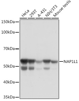
Western blot analysis of various lysates using NAP1L1 Rabbit mAb (CAB6174) at 1:1000 dilution. Secondary antibody: HRP-conjugated Goat anti-Rabbit IgG (H+L) (CABS014) at 1:10000 dilution. Lysates/proteins: 25μg per lane. Blocking buffer: 3% nonfat dry milk in TBST. Detection: ECL Basic Kit (AbGn00020). Exposure time: 10s.
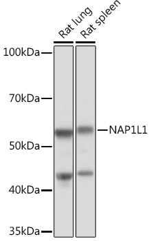
Western blot analysis of various lysates using NAP1L1 Rabbit mAb (CAB6174) at 1:1000 dilution. Secondary antibody: HRP-conjugated Goat anti-Rabbit IgG (H+L) (CABS014) at 1:10000 dilution. Lysates/proteins: 25μg per lane. Blocking buffer: 3% nonfat dry milk in TBST. Detection: ECL Basic Kit (AbGn00020). Exposure time: 1min.
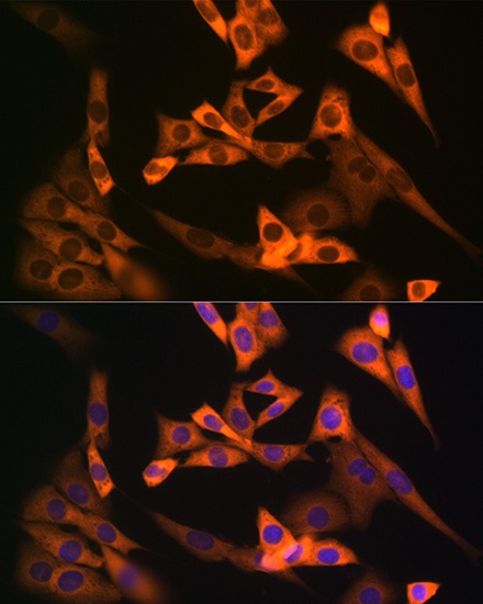
Immunofluorescence analysis of NIH-3T3 cells using NAP1L1 Rabbit mAb (CAB6174) at dilution of 1:100 (40x lens). Secondary antibody: Cy3-conjugated Goat anti-Rabbit IgG (H+L) (CABS007) at 1:500 dilution. Blue: DAPI for nuclear staining.
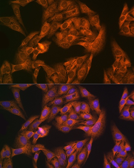
Immunofluorescence analysis of U-2 OS cells using NAP1L1 Rabbit mAb (CAB6174) at dilution of 1:100 (40x lens). Secondary antibody: Cy3-conjugated Goat anti-Rabbit IgG (H+L) (CABS007) at 1:500 dilution. Blue: DAPI for nuclear staining.