The NAT10 Monoclonal Antibody (CAB19286) is a high-quality antibody developed for reliable detection and analysis of target proteins. This antibody, raised in rabbits, is highly specific to human samples and has been validated for use in Western blot applications. By binding to NAT10, the antibody allows for accurate detection and analysis of this protein in a variety of cell types, making it an essential tool for studies in molecular biology and cancer research.NAT10 is an essential enzyme involved in the maturation of ribosomal RNA and the regulation of cell growth, making it a key player in processes like protein synthesis and cell proliferation.
This antibody is validated for use in WB, IHC-P, ELISA applications and has demonstrated reactivity against Human, Mouse, Rat samples.
Product Name:
NAT10 Monoclonal Antibody
SKU:
CAB19286
Size:
20μL, 100μL
Reactivity:
Human, Mouse, Rat
Clone Number:
ARC2468
Conjugate:
Unconjugated
Immunogen:
Recombinant protein (or fragment).This information is considered to be commercially sensitive.
Recommended starting concentration is 1 μg/mL. Please optimize the concentration based on your specific assay requirements.
Synonyms:
ALP, Kre33, NET43, NAT10
Positive Sample:
HeLa, SH-SY5Y, Mouse testis
Cellular Localization:
Nucleus, Nucleolus.
Calculated MW:
116kDa
Observed MW:
120kDa
The protein encoded by this gene is an RNA cytidine acetyltransferase involved in histone acetylation, tRNA acetylation, the biosynthesis of 18S rRNA, and the enhancement of nuclear architecture and chromatin organization.
Purification Method
Affinity purification
Gene ID
55226
Buffer Information
Store at -20℃. Avoid freeze / thaw cycles. Buffer: PBS containing 50% glycerol and 0.05% BSA, preserved with proclin300 or sodium azide, pH 7.3.
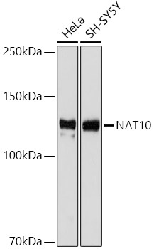
Western blot analysis of various lysates using NAT10 Rabbit mAb (CAB19286) at 1:1000 dilution. Secondary antibody: HRP-conjugated Goat anti-Rabbit IgG (H+L) (CABS014) at 1:10000 dilution. Lysates/proteins: 25μg per lane. Blocking buffer: 3% nonfat dry milk in TBST. Detection: ECL Basic Kit (AbGn00020). Exposure time: 3s.
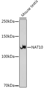
Western blot analysis of lysates from Mouse testis, using NAT10 Rabbit mAb (CAB19286) at 1:1000 dilution. Secondary antibody: HRP-conjugated Goat anti-Rabbit IgG (H+L) (CABS014) at 1:10000 dilution. Lysates/proteins: 25μg per lane. Blocking buffer: 3% nonfat dry milk in TBST. Detection: ECL Basic Kit (AbGn00020). Exposure time: 30s.
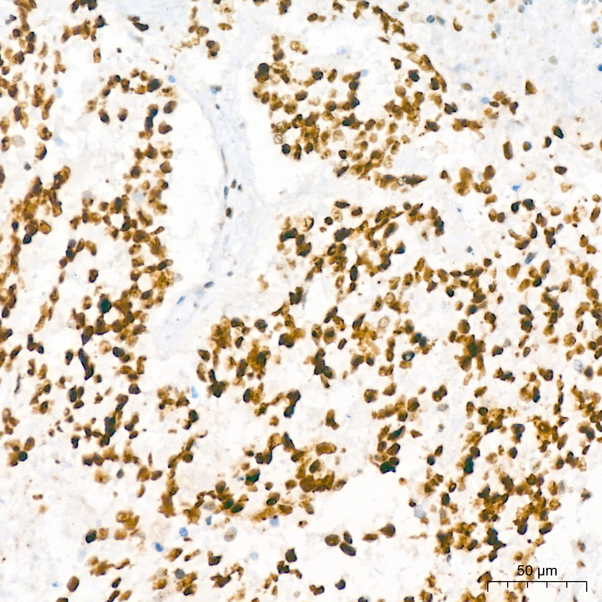
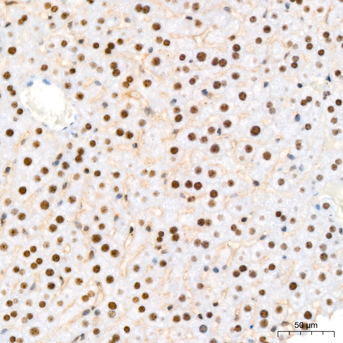
Immunohistochemistry analysis of paraffin-embedded Human colon using NAT10 Rabbit mAb (CAB19286) at dilution of 1:200 (40x lens). High pressure antigen retrieval performed with 0.01M Tris/EDTA Buffer (pH 9.0) prior to IHC staining.
Immunohistochemistry analysis of paraffin-embedded Human lung squamous carcinoma tissue using NAT10 Rabbit mAb (CAB19286) at dilution of 1:200 (40x lens). High pressure antigen retrieval performed with 0.01M Tris/EDTA Buffer (pH 9.0) prior to IHC staining.
Immunohistochemistry analysis of paraffin-embedded Mouse liver using NAT10 Rabbit mAb (CAB19286) at dilution of 1:200 (40x lens). High pressure antigen retrieval performed with 0.01M Tris/EDTA Buffer (pH 9.0) prior to IHC staining.
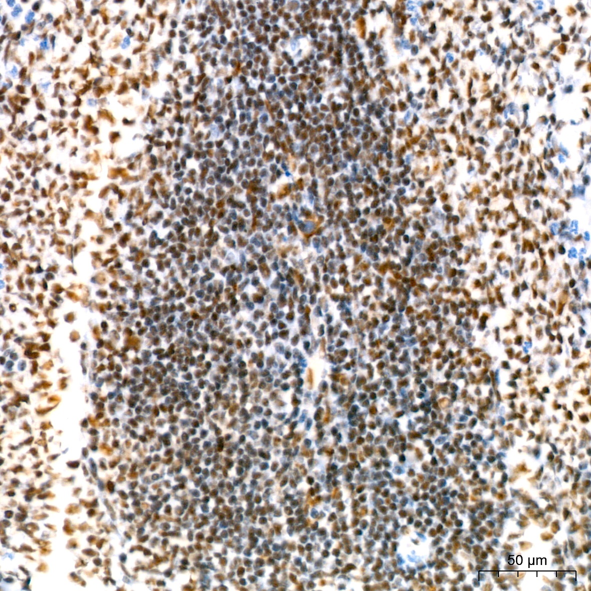
Immunohistochemistry analysis of paraffin-embedded Rat spleen using NAT10 Rabbit mAb (CAB19286) at dilution of 1:200 (40x lens). High pressure antigen retrieval performed with 0.01M Tris/EDTA Buffer (pH 9.0) prior to IHC staining.