The NLK Monoclonal Antibody (CAB19270) is a high-quality antibody developed for reliable detection and analysis of target proteins. This antibody, developed through rabbit immunization, demonstrates high reactivity with human samples and is suitable for Western blot applications.NLK, also known as Nemo-like kinase, is a critical component in signal transduction pathways that govern various cellular functions. Its involvement in pathways related to cancer, developmental biology, and inflammation make it an attractive target for further investigation.
This antibody is validated for use in WB, IHC-P, ELISA applications and has demonstrated reactivity against Human samples.
Product Name:
NLK Monoclonal Antibody
SKU:
CAB19270
Size:
20μL, 100μL
Reactivity:
Human
Clone Number:
ARC2441
Conjugate:
Unconjugated
Immunogen:
Synthetic peptide. This information is considered to be commercially sensitive.
Recommended starting concentration is 1 μg/mL. Please optimize the concentration based on your specific assay requirements.
Synonyms:
NLK, nemo like kinase, LAK1
Positive Sample:
HeLa, SW480
Cellular Localization:
Cytoplasm, Nucleus.
Calculated MW:
58kDa
Observed MW:
58kDa
Enables ubiquitin protein ligase binding activity. Involved in protein stabilization and transforming growth factor beta receptor signaling pathway. Predicted to be located in cytosol and nucleoplasm. Predicted to be active in cytoplasm and nucleus.
Purification Method
Affinity purification
Gene ID
51701
Buffer Information
Store at -20℃. Avoid freeze / thaw cycles. Buffer: PBS containing 50% glycerol and 0.05% BSA, preserved with proclin300 or sodium azide, pH 7.3.
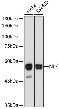
Western blot analysis of various lysates using NLK Rabbit mAb (CAB19270) at 1:1000 dilution. Secondary antibody: HRP-conjugated Goat anti-Rabbit IgG (H+L) (CABS014) at 1:10000 dilution. Lysates/proteins: 25μg per lane. Blocking buffer: 3% nonfat dry milk in TBST. Detection: ECL Basic Kit (AbGn00020). Exposure time: 180s.
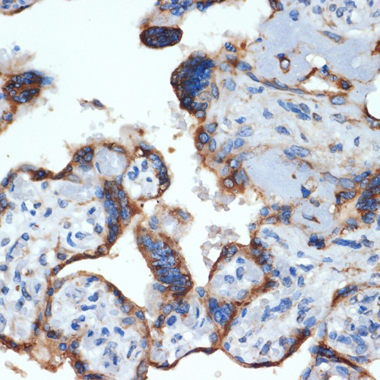
Immunohistochemistry analysis of paraffin-embedded Human placenta using NLK Rabbit mAb (CAB19270) at dilution of 1:100 (40x lens). Microwave antigen retrieval performed with 0.01M Tris/EDTA Buffer (pH 9.0) prior to IHC staining.