The PCNT Antibody (CAB20161) is a high-quality antibody developed for reliable detection and analysis of target proteins. This antibody, produced in rabbits, is highly specific to human samples and is validated for use in various applications, including immunofluorescence, immunohistochemistry, and Western blot.Pericentrin is a key player in centrosome function, having implications in cell cycle progression, microtubule organization, and cell division accuracy. Dysregulation of PCNT has been linked to various human diseases, including cancer and neurological disorders.
This antibody is validated for use in WB, IF/ICC, ELISA applications and has demonstrated reactivity against Human, Rat samples.
Product Name:
PCNT Antibody
SKU:
CAB20161
Size:
20μL, 100μL
Reactivity:
Human, Rat
Conjugate:
Unconjugated
Immunogen:
Recombinant protein (or fragment).This information is considered to be commercially sensitive.
Recommended starting concentration is 1 μg/mL. Please optimize the concentration based on your specific assay requirements.
Synonyms:
KEN, PCN, MOPD2, PCNT2, PCNTB, PCTN2, SCKL4, PCNT
Positive Sample:
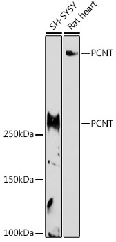
SH-SY5Y, Rat heart
Cellular Localization:
Centriole, Centrosome, Cytosol.
Calculated MW:
378kDa
Observed MW:
251kDa/378kDa
The protein encoded by this gene binds to calmodulin and is expressed in the centrosome. It is an integral component of the pericentriolar material (PCM). The protein contains a series of coiled-coil domains and a highly conserved PCM targeting motif called the PACT domain near its C-terminus. The protein interacts with the microtubule nucleation component gamma-tubulin and is likely important to normal functioning of the centrosomes, cytoskeleton, and cell-cycle progression. Mutations in this gene cause Seckel syndrome-4 and microcephalic osteodysplastic primordial dwarfism type II. Two transcript variants encoding different isoforms have been found for this gene.
Purification Method
Affinity purification
Gene ID
5116
RRID
AB_2862948
Buffer Information
Store at -20℃. Avoid freeze / thaw cycles. Buffer: PBS containing 50% glycerol, preserved with proclin300 or sodium azide, pH 7.3.
Western blot analysis of various lysates using PCNT Rabbit pAb (CAB20161) at 1:1000 dilution. Secondary antibody: HRP-conjugated Goat anti-Rabbit IgG (H+L) (CABS014) at 1:10000 dilution. Lysates/proteins: 25μg per lane. Blocking buffer: 3% nonfat dry milk in TBST. Detection: ECL Basic Kit (AbGn00020). Exposure time: 180s.
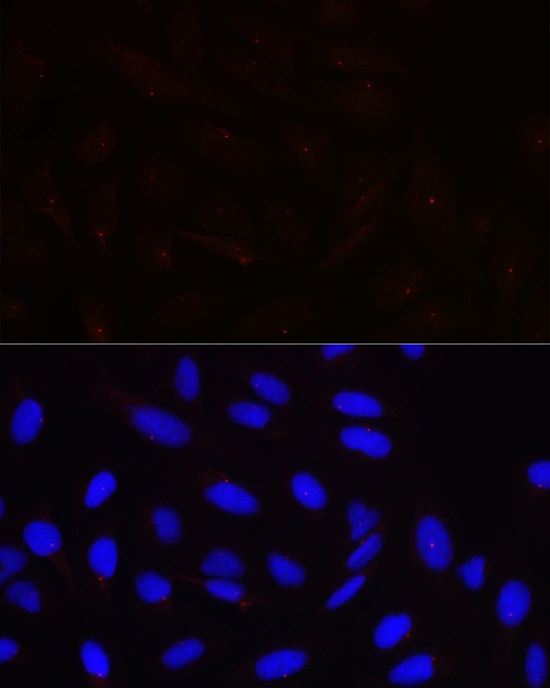
Immunofluorescence analysis of U-2 OS cells using PCNT Rabbit pAb (CAB20161) at dilution of 1:100 (40x lens). Secondary antibody: Cy3-conjugated Goat anti-Rabbit IgG (H+L) (CABS007) at 1:500 dilution. Blue: DAPI for nuclear staining.