The PDCD7 Monoclonal Antibody (CAB1510) is a high-quality antibody developed for reliable detection and analysis of target proteins. This antibody, produced in rabbits, has high specificity for human samples and is validated for use in Western blot applications. By targeting the PDCD7 protein, researchers can examine its role in various cellular processes and gain insights into its function in physiology and disease.PDCD7 is a key player in programmed cell death and cellular stress responses, making it a promising target for studies in cancer biology, developmental biology, and genomics.
This antibody is validated for use in WB, IHC-P, ELISA applications and has demonstrated reactivity against Human, Mouse, Rat samples.
Product Name:
PDCD7 Monoclonal Antibody
SKU:
CAB1510
Size:
20μL, 100μL
Reactivity:
Human, Mouse, Rat
Clone Number:
ARC2564
Conjugate:
Unconjugated
Immunogen:
Synthetic peptide. This information is considered to be commercially sensitive.
Recommended starting concentration is 1 μg/mL. Please optimize the concentration based on your specific assay requirements.
Synonyms:
59K, ES18, HES18, PDCD7
Positive Sample:
TE-1, SK-OV-3, Mouse testis, Mouse thymus, Rat brain
Cellular Localization:
Nucleus.
Calculated MW:
55kDa
Observed MW:
55kDa
This gene encodes a 59 kDa protein that is associated with the U11 small nuclear ribonucleoprotein (snRNP), which is a component of the minor U12-type spliceosome responsible for catalyzing pre-mRNA splicing of U12-type introns.
Purification Method
Affinity purification
Gene ID
10081
Buffer Information
Store at -20℃. Avoid freeze / thaw cycles. Buffer: PBS containing 50% glycerol and 0.05% BSA, preserved with proclin300 or sodium azide, pH 7.3.
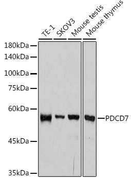
Western blot analysis of various lysates using PDCD7 Rabbit mAb (CAB1510) at 1:1000 dilution. Secondary antibody: HRP-conjugated Goat anti-Rabbit IgG (H+L) (CABS014) at 1:10000 dilution. Lysates/proteins: 25μg per lane. Blocking buffer: 3% nonfat dry milk in TBST. Detection: ECL Basic Kit (AbGn00020). Exposure time: 10s.
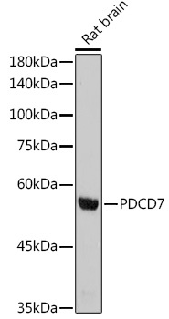
Western blot analysis of lysates from Rat brain, using PDCD7 Rabbit mAb (CAB1510) at 1:1000 dilution. Secondary antibody: HRP-conjugated Goat anti-Rabbit IgG (H+L) (CABS014) at 1:10000 dilution. Lysates/proteins: 25μg per lane. Blocking buffer: 3% nonfat dry milk in TBST. Detection: ECL Basic Kit (AbGn00020). Exposure time: 90s.
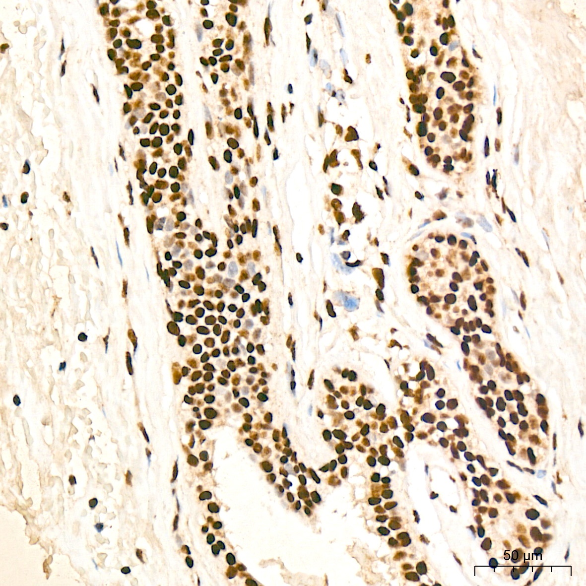
Immunohistochemistry analysis of paraffin-embedded Human breast tissue using PDCD7 Rabbit mAb (CAB1510) at a dilution of 1:200 (40x lens). High pressure antigen retrieval was performed with 0.01 M citrate buffer (pH 6.0) prior to IHC staining.
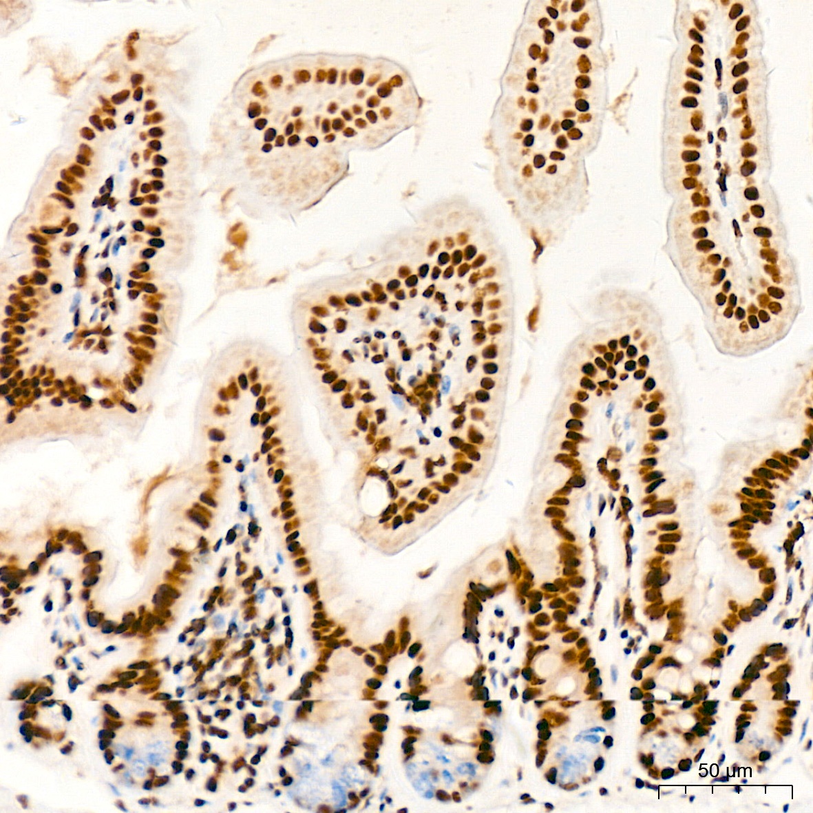
Immunohistochemistry analysis of paraffin-embedded Human small intestine tissue using PDCD7 Rabbit mAb (CAB1510) at a dilution of 1:200 (40x lens). High pressure antigen retrieval was performed with 0.01 M citrate buffer (pH 6.0) prior to IHC staining.
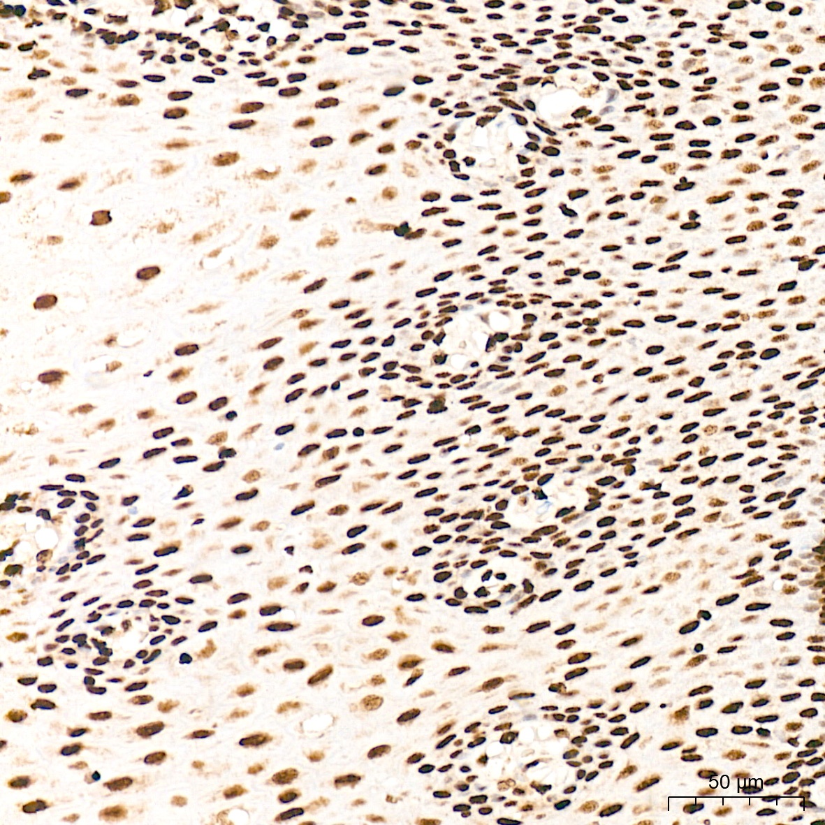
Immunohistochemistry analysis of paraffin-embedded Human esophagus tissue using PDCD7 Rabbit mAb (CAB1510) at a dilution of 1:200 (40x lens). High pressure antigen retrieval was performed with 0.01 M citrate buffer (pH 6.0) prior to IHC staining.
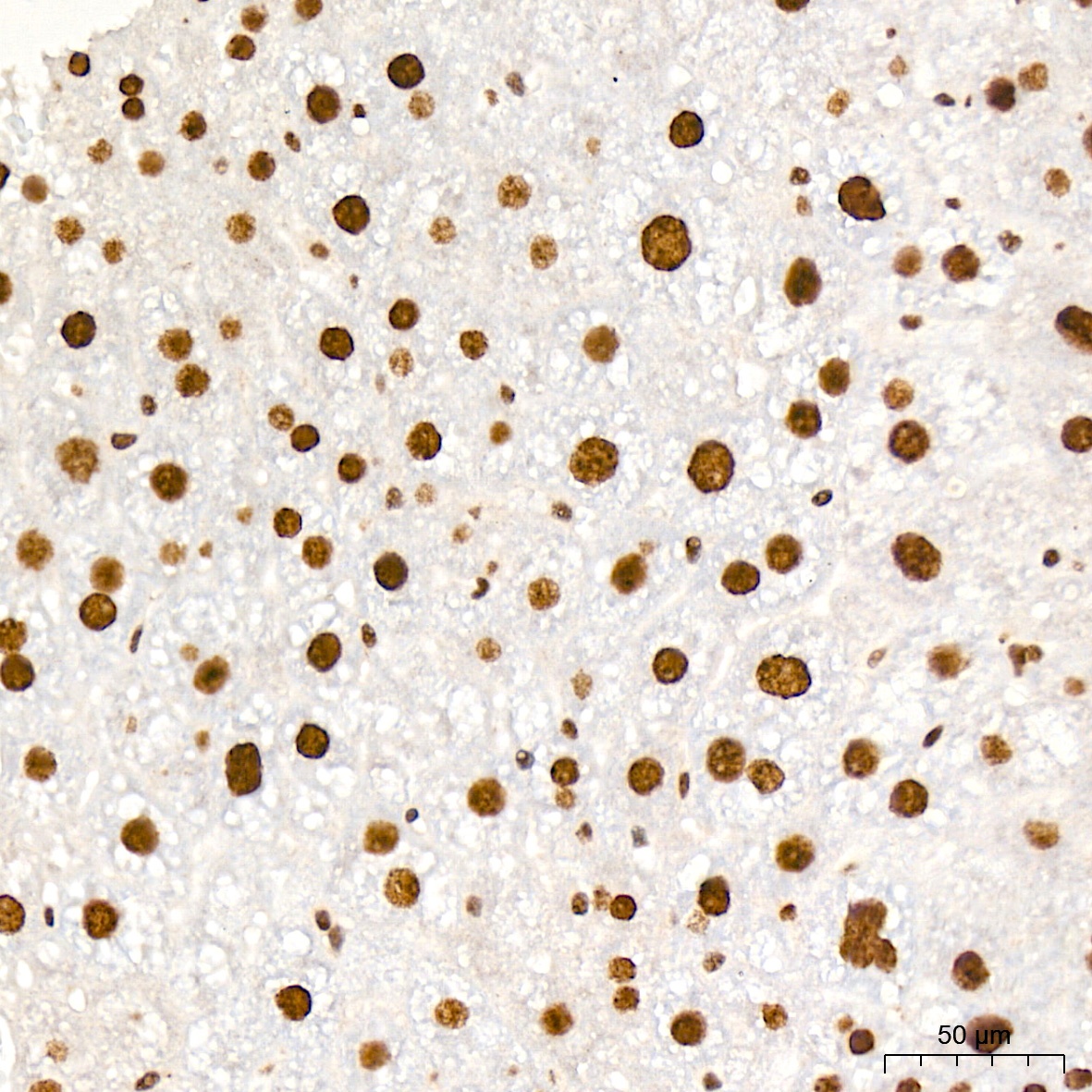
Immunohistochemistry analysis of paraffin-embedded Rat liver tissue using PDCD7 Rabbit mAb (CAB1510) at a dilution of 1:200 (40x lens). High pressure antigen retrieval was performed with 0.01 M citrate buffer (pH 6.0) prior to IHC staining.