The Phospho-BRD4-S1070 Antibody (CABP1181) is a high-quality antibody developed for reliable detection and analysis of target proteins. This antibody, produced in rabbits, specifically detects phosphorylation at serine 1070 in BRD4 and is validated for use in Western blot and immunofluorescence applications.Phosphorylation of BRD4 at serine 1070 has been linked to its role in controlling transcription and cell cycle progression, highlighting its importance in various biological processes. By targeting this specific phosphorylation site, researchers can gain insight into the regulatory mechanisms involving BRD4 and its impact on cellular functions.
This antibody is validated for use in WB, ELISA applications and has demonstrated reactivity against Human samples.
Product Name:
Phospho-BRD4-S1070 Antibody
SKU:
CABP1181
Size:
20μL, 100μL
Reactivity:
Human
Immunogen:
Synthetic peptide. This information is considered to be commercially sensitive.
Sequence:
IHSP Q
Tested Applications:
WBELISA
Recommended Dilution:
WB
1:500 - 1:1000
ELISA
Recommended starting concentration is 1 μg/mL. Please optimize the concentration based on your specific assay requirements.
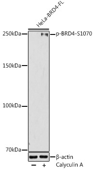
HeLa treated with Calyculin A after transfected with BRD4 (Full length)
Cellular Localization:
Chromosome, Nucleus.
Calculated MW:
152kDa
Observed MW:
250kDa
The protein encoded by this gene is homologous to the murine protein MCAP, which associates with chromosomes during mitosis, and to the human RING3 protein, a serine/threonine kinase. Each of these proteins contains two bromodomains, a conserved sequence motif which may be involved in chromatin targeting. This gene has been implicated as the chromosome 19 target of translocation t(15;19)(q13;p13.1), which defines an upper respiratory tract carcinoma in young people. Two alternatively spliced transcript variants have been described.
Purification Method
Affinity purification
Gene ID
23476
RRID
AB_2864039
Buffer Information
Store at -20℃. Avoid freeze / thaw cycles. Buffer: PBS containing 50% glycerol, preserved with proclin300 or sodium azide, pH 7.3.
Western blot analysis of lysates from HeLa cells, using Phospho-BRD4-S1070 Rabbit pAb (CABP1181) at 1:1000 dilution. HeLa cells were treated with Calyculin A (100 nM) at 37℃ for 30 minutes after serum-starvation overnight. Secondary antibody: HRP-conjugated Goat anti-Rabbit IgG (H+L) (CABS014) at 1:10000 dilution. Lysates/proteins: 25μg per lane. Blocking buffer: 3% nonfat dry milk in TBST. Detection: ECL Enhanced Kit (AbGn00021). Exposure time: 180s.


")
