The Phospho-c-Jun-T91 Antibody (CABP1216) is a high-quality antibody developed for reliable detection and analysis of target proteins. This polyclonal antibody, generated in rabbits, has been validated for use in Western blot applications and is highly reactive with human samples.Phosphorylation of c-Jun at Threonine 91 is known to play a crucial role in the activation of c-Jun, influencing its ability to regulate gene expression and participate in various cellular processes such as proliferation, differentiation, and apoptosis. The Anti-Phospho c-Jun (T91) Antibody (CABP1216) specifically binds to the phosphorylated form of c-Jun at Threonine 91, enabling researchers to detect and analyze its activity in different cell types.
This antibody is validated for use in WB, IHC-P, IF/ICC, ELISA applications and has demonstrated reactivity against Human, Mouse, Rat samples.
Product Name:
Phospho-c-Jun-T91 Antibody
SKU:
CABP1216
Size:
20μL, 100μL
Reactivity:
Human, Mouse, Rat
Conjugate:
Unconjugated
Immunogen:
Synthetic peptide. This information is considered to be commercially sensitive.
Sequence:
TTTP T
Tested Applications:
WBIHC-PIF/ICCELISA
Recommended Dilution:
WB
1:500 - 1:1000
IHC-P
1:50 - 1:200
IF/ICC
1:50 - 1:200
ELISA
Recommended starting concentration is 1 μg/mL. Please optimize the concentration based on your specific assay requirements.
Synonyms:
AP1, p39, AP-1, cJUN, c-Jun, Phospho-c-Jun-T91
Positive Sample:
293T treated with Anisomycin, 293T treated with UV, NIH/3T3 treated with Anisomycin, NIH/3T3 treated with UV
Cellular Localization:
Nucleus.
Calculated MW:
36kDa
Observed MW:
36kDa
This gene is the putative transforming gene of avian sarcoma virus 17. It encodes a protein which is highly similar to the viral protein, and which interacts directly with specific target DNA sequences to regulate gene expression. This gene is intronless and is mapped to 1p32-p31, a chromosomal region involved in both translocations and deletions in human malignancies.
Purification Method
Affinity purification
Gene ID
3725
Buffer Information
Store at -20℃. Avoid freeze / thaw cycles. Buffer: PBS containing 50% glycerol, preserved with proclin300 or sodium azide, pH 7.3.
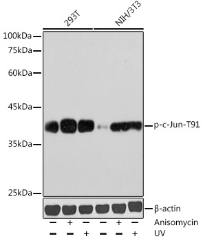
Western blot analysis of various lysates using Phospho-c-Jun-T91 Rabbit pAb (CABP1216) at 1:1000 dilution. Both 293T cells and NIH/3T3 cells were treated with Anisomycin (25 μg/mL) at 37℃ for 30 minutes after serum-starvation overnight. Both 293T cells and NIH/3T3 cells were treated with UV at room temperature for 15-30 minutes. Secondary antibody: HRP-conjugated Goat anti-Rabbit IgG (H+L) (CABS014) at 1:10000 dilution. Lysates/proteins: 25μg per lane. Blocking buffer: 3% nonfat dry milk in TBST. Detection: ECL Basic Kit (AbGn00020). Exposure time: 180s.
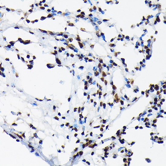
Immunohistochemistry analysis of paraffin-embedded Rat lung using Phospho-c-Jun-T91 Rabbit pAb (CABP1216) at dilution of 1:50 (40x lens). High pressure antigen retrieval performed with 0.01M Citrate buffer (pH 6.0) prior to IHC staining.
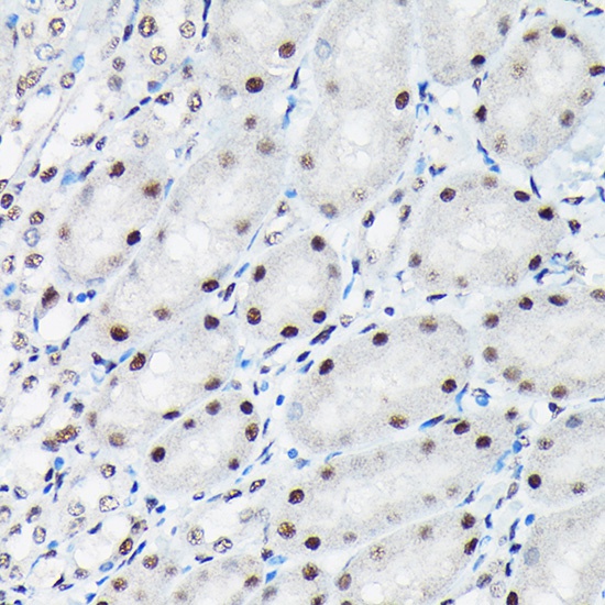
Immunohistochemistry analysis of paraffin-embedded Mouse kidney using Phospho-c-Jun-T91 Rabbit pAb (CABP1216) at dilution of 1:50 (40x lens). High pressure antigen retrieval performed with 0.01M Citrate buffer (pH 6.0) prior to IHC staining.
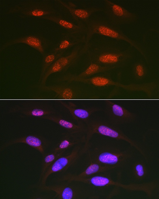
Immunofluorescence analysis of U2OS cells using Phospho-c-Jun-T91 Rabbit pAb (CABP1216) at dilution of 1:50 (40x lens). Secondary antibody: Cy3-conjugated Goat anti-Rabbit IgG (H+L) (CABS007) at 1:500 dilution. Blue: DAPI for nuclear staining.