The Phospho-Cdc6-S54 Monoclonal Antibody (CABP1153) is a high-quality antibody developed for reliable detection and analysis of target proteins. The protein encoded by this gene is highly similar to Saccharomyces cerevisiae Cdc6, a protein essential for the initiation of DNA replication. This protein functions as a regulator at the early steps of DNA replication. It localizes in cell nucleus during cell cyle G1, but translocates to the cytoplasm at the start of S phase. The subcellular translocation of this protein during cell cyle is regulated through its phosphorylation by Cdks. Transcription of this protein was reported to be regulated in response to mitogenic signals through transcriptional control mechanism involving E2F proteins.
This antibody is validated for use in WB, ELISA applications and has demonstrated reactivity against Mouse samples.
Product Name:
Phospho-Cdc6-S54 Monoclonal Antibody
SKU:
CABP1153
Size:
100μL, 20μL
Reactivity:
Mouse
Clone Number:
ARC1693
Conjugate:
Unconjugated
Immunogen:
Synthetic peptide. This information is considered to be commercially sensitive.
Tested Applications:
WBELISA
Recommended Dilution:
WB
1:1000 - 1:6000
ELISA
Recommended starting concentration is 1 μg/mL. Please optimize the concentration based on your specific assay requirements.
Synonyms:
CDC18L, HsCDC6, MGORS5, HsCDC18, Phospho-Cdc6-S54
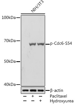
Positive Sample:
NIH/3T3 treated with Paclitaxel, NIH/3T3 treated with Hydroxyurea
Cellular Localization:
Cytoplasm, Nucleus.
Calculated MW:
63kDa
Observed MW:
63kDa
The protein encoded by this gene is highly similar to Saccharomyces cerevisiae Cdc6, a protein essential for the initiation of DNA replication. This protein functions as a regulator at the early steps of DNA replication. It localizes in cell nucleus during cell cyle G1, but translocates to the cytoplasm at the start of S phase. The subcellular translocation of this protein during cell cyle is regulated through its phosphorylation by Cdks. Transcription of this protein was reported to be regulated in response to mitogenic signals through transcriptional control mechanism involving E2F proteins.
Purification Method
Affinity purification
Gene ID
990
RRID
AB_2864017
Buffer Information
Store at -20℃. Avoid freeze / thaw cycles. Buffer: PBS containing 50% glycerol and 0.05% BSA, preserved with proclin300 or sodium azide, pH 7.3.
Western blot analysis of lysates from NIH/3T3 cells, using Phospho-Cdc6-S54 Rabbit mAb (CABP1153) at 1:1000 dilution. NIH/3T3 cells were treated with Paclitaxel (1 uM) at 37℃ for 20 hours. NIH/3T3 cells were treated with Hydroxyurea (4 mM) at 37℃ for 20 hours. Secondary antibody: HRP-conjugated Goat anti-Rabbit IgG (H+L) (AS014) at 1:10000 dilution. Lysates/proteins: 25μg per lane. Blocking buffer: 3% BSA. Detection: ECL Basic Kit (AbGn00020). Exposure time: 5s.