The Phospho-IkappaBalpha-S32 Monoclonal Antibody (CABP0707) is a high-quality antibody developed for reliable detection and analysis of target proteins. This highly specific antibody, produced in rabbits, is validated for use in Western blot applications and is particularly reactive with human samples.Phosphorylation of IKBalpha at S32 plays a critical role in the regulation of NF-kB activity, making this antibody essential for studies aiming to understand the intricacies of immune signaling and the development of targeted therapies for inflammatory disorders, autoimmune diseases, and cancer.
This antibody is validated for use in WB, ELISA applications and has demonstrated reactivity against Human, Mouse, Rat samples.
Product Name:
Phospho-IkappaBalpha-S32 Monoclonal Antibody
SKU:
CABP0707
Size:
20μL, 100μL
Reactivity:
Human, Mouse, Rat
Clone Number:
ARC0147
Conjugate:
Unconjugated
Immunogen:
Synthetic peptide. This information is considered to be commercially sensitive.
Sequence:
HDSG L
Tested Applications:
WBELISA
Recommended Dilution:
WB
1:500 - 1:2000
ELISA
Recommended starting concentration is 1 μg/mL. Please optimize the concentration based on your specific assay requirements.
Synonyms:
IKBA, MAD-3, NFKBI, EDAID2, Phospho-IκBα-S32
Positive Sample:
NIH/3T3 treated with TNF-α, C6
Cellular Localization:
Cytoplasm, Nucleus.
Calculated MW:
36kDa
Observed MW:
39kDa
This gene encodes a member of the NF-kappa-B inhibitor family, which contain multiple ankrin repeat domains. The encoded protein interacts with REL dimers to inhibit NF-kappa-B/REL complexes which are involved in inflammatory responses. The encoded protein moves between the cytoplasm and the nucleus via a nuclear localization signal and CRM1-mediated nuclear export. Mutations in this gene have been found in ectodermal dysplasia anhidrotic with T-cell immunodeficiency autosomal dominant disease.
Purification Method
Affinity purification
Gene ID
4792
RRID
AB_2863811
Buffer Information
Store at -20℃. Avoid freeze / thaw cycles. Buffer: PBS containing 50% glycerol and 0.05% BSA, preserved with proclin300 or sodium azide, pH 7.3.
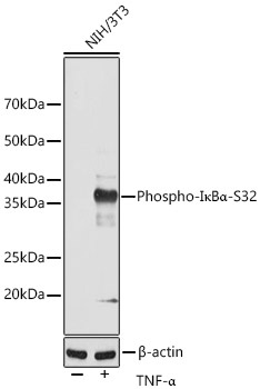
Western blot analysis of lysates from NIH/3T3 cells, using Phospho-IκBα-S32 Rabbit mAb (CABP0707) at 1:1000 dilution. NIH/3T3 and C6 cells were treated with TNF-α (20 ng/mL) at 37℃ for 30 minutes. Secondary antibody: HRP-conjugated Goat anti-Rabbit IgG (H+L) (CABS014) at 1:10000 dilution. Lysates/proteins: 25μg per lane. Blocking buffer: 3% nonfat dry milk in TBST. Detection: ECL Basic Kit (AbGn00020). Exposure time: 120s.
Western blot analysis of lysates from C6 cells using Phospho-IκBα-S32 Rabbit mAb (CABP0707) at 1:1000 dilution (upper) or IκBα Rabbit mAb (CAB24909) at1:6000 dilution (lower) incubated overnight at 4℃. C6 cells were treated with Calyculin A (100 nM) at 37℃ for 30 minutes after serum-starvation overnight. Secondary antibody: HRP-conjugated Goat anti-Rabbit IgG (H+L) (CABS014) at 1:10000 dilution. Lysates/proteins: 30 μg per lane. Blocking buffer: 3 % nonfat dry milk in TBST. Detection: ECL Basic Kit (AbGn00020). Exposure time: 60s.