The Phospho-IkappaBalpha-S36 Monoclonal Antibody (CABP0999) is a high-quality antibody developed for reliable detection and analysis of target proteins. This antibody, generated in rabbits, specifically recognizes the phosphorylated form of IκBα at serine 36 in human samples and is validated for use in Western blot applications. By targeting this phosphorylation site, researchers can gain insight into the regulation of NF-κB signaling and its involvement in various physiological and pathological processes.The NF-κB signaling pathway is a key regulator of immune responses, inflammation, and cell survival, making it a focus of research in immunology, cancer biology, and inflammatory diseases.
This antibody is validated for use in WB, IHC-P, ELISA applications and has demonstrated reactivity against Human, Mouse, Rat samples.
Product Name:
Phospho-IkappaBalpha-S36 Monoclonal Antibody
SKU:
CABP0999
Size:
20μL, 100μL
Reactivity:
Human, Mouse, Rat
Clone Number:
ARC1543
Conjugate:
Unconjugated
Immunogen:
Synthetic peptide. This information is considered to be commercially sensitive.
Sequence:
LDSM K
Tested Applications:
WBIHC-PELISA
Recommended Dilution:
WB
1:500 - 1:1000
IHC-P
1:50 - 1:200
ELISA
Recommended starting concentration is 1 μg/mL. Please optimize the concentration based on your specific assay requirements.
Synonyms:
IKBA, MAD-3, NFKBI, EDAID2, Phospho-IκBα-S36
Positive Sample:
THP-1 treated with LPS, THP-1 treated with PMA, NIH/3T3 treated with TNF-α, C6 treated with TNF-α
Cellular Localization:
Cytoplasm, Nucleus.
Calculated MW:
36kDa
Observed MW:
40kDa
This gene encodes a member of the NF-kappa-B inhibitor family, which contain multiple ankrin repeat domains. The encoded protein interacts with REL dimers to inhibit NF-kappa-B/REL complexes which are involved in inflammatory responses. The encoded protein moves between the cytoplasm and the nucleus via a nuclear localization signal and CRM1-mediated nuclear export. Mutations in this gene have been found in ectodermal dysplasia anhidrotic with T-cell immunodeficiency autosomal dominant disease.
Purification Method
Affinity purification
Gene ID
4792
RRID
AB_2863891
Buffer Information
Store at -20℃. Avoid freeze / thaw cycles. Buffer: PBS containing 50% glycerol and 0.05% BSA, preserved with proclin300 or sodium azide, pH 7.3.
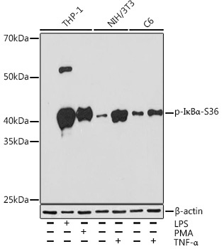
Western blot analysis of various lysates using Phospho-IκBα-S36 Rabbit mAb (CABP0999) at 1:1000 dilution. THP-1 cells were treated with PMA/TPA (80 nM) at 37℃ for overnight or treated with LPS (1 μg/mL) at 37℃ for 6 hours. Both NIH/3T3 cells and C6 cells were treated with TNF-α (20 ng/mL) at 37℃ for 30 minutes. Secondary antibody: HRP-conjugated Goat anti-Rabbit IgG (H+L) (CABS014) at 1:10000 dilution. Lysates/proteins: 25μg per lane. Blocking buffer: 3% BSA. Detection: ECL Enhanced Kit (AbGn00021). Exposure time: 3min.
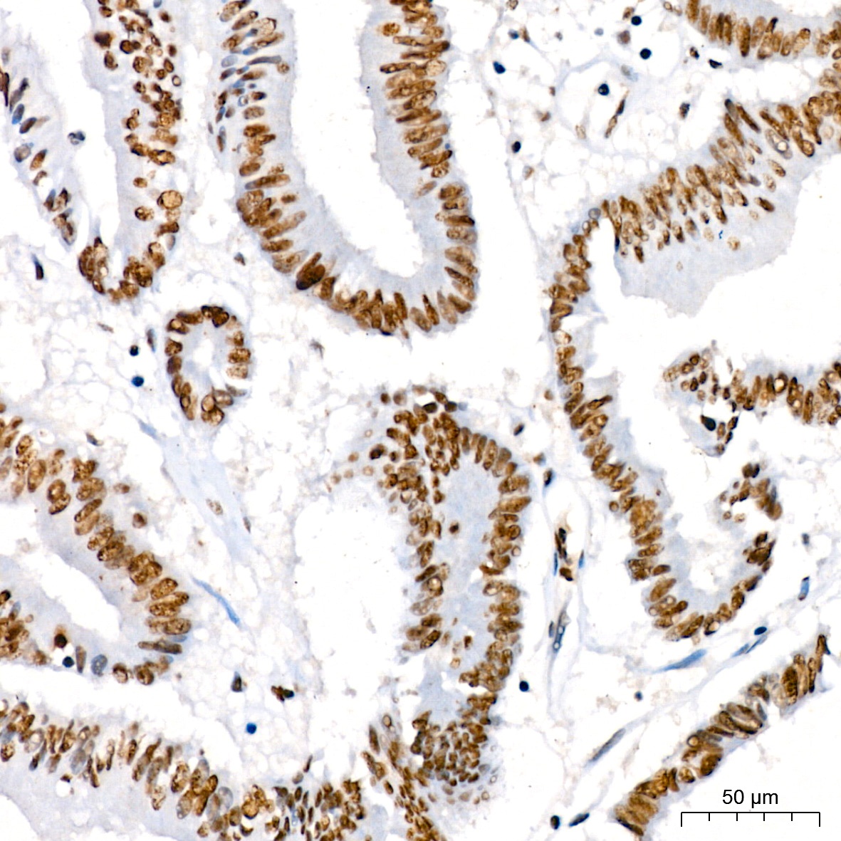
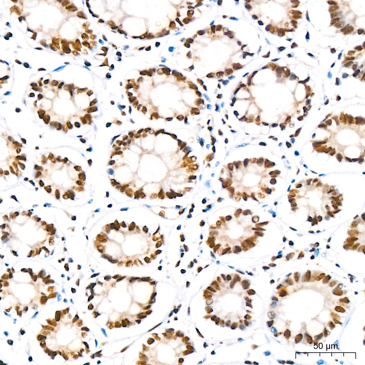
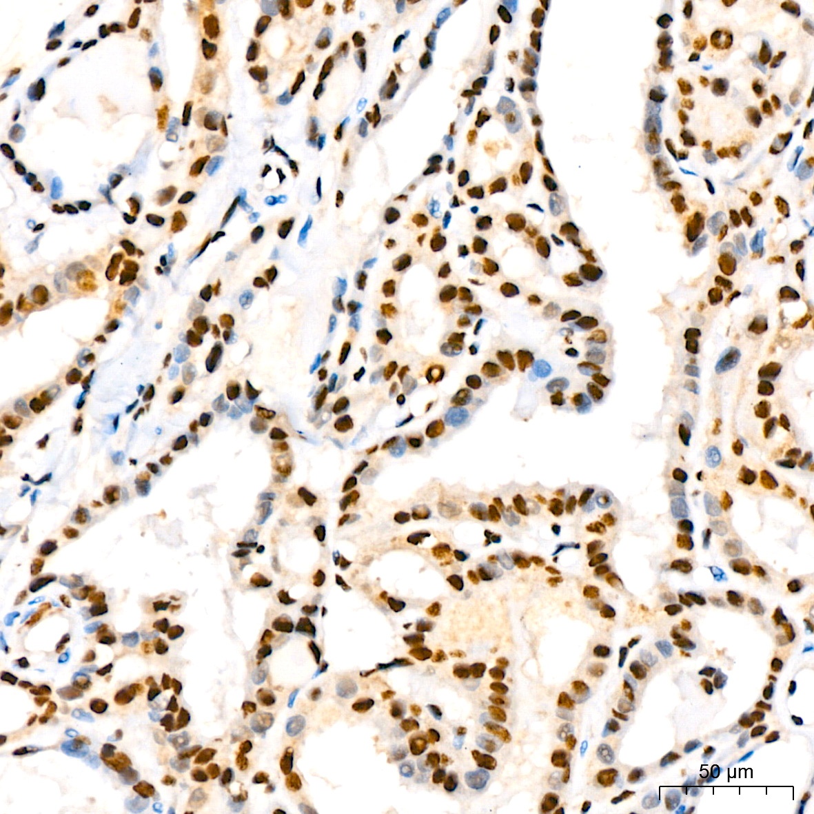
Immunohistochemistry analysis of paraffin-embedded Human colon carcinoma using Phospho-IκBα-S36 Rabbit mAb (CABP0999) at dilution of 1:200 (40x lens). High pressure antigen retrieval performed with 0.01M Citrate buffer (pH 6.0) prior to IHC staining.
Immunohistochemistry analysis of paraffin-embedded Human colon using Phospho-IκBα-S36 Rabbit mAb (CABP0999) at dilution of 1:200 (40x lens). High pressure antigen retrieval performed with 0.01M Citrate buffer (pH 6.0) prior to IHC staining.
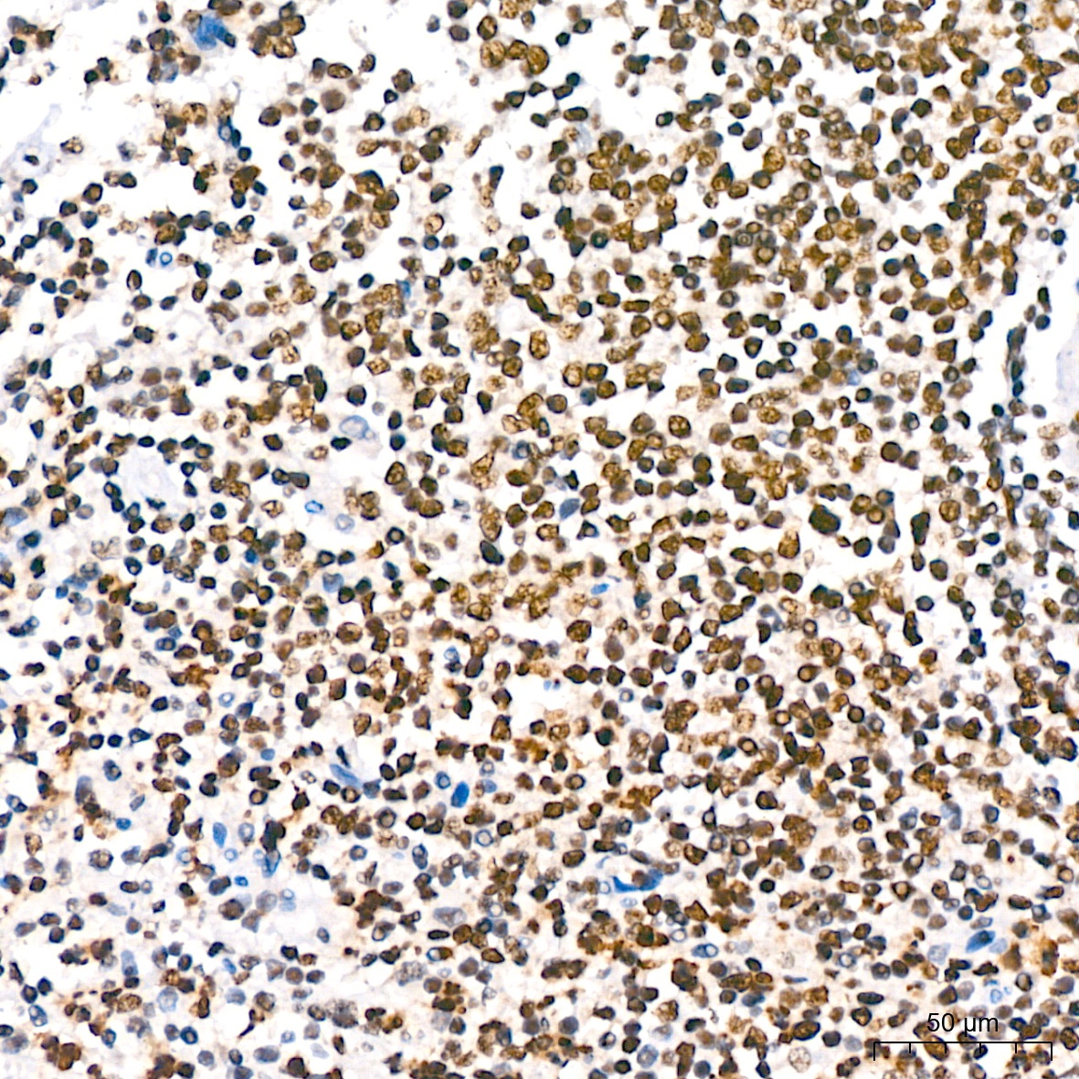
Immunohistochemistry analysis of paraffin-embedded Human spleen using Phospho-IκBα-S36 Rabbit mAb (CABP0999) at dilution of 1:200 (40x lens). High pressure antigen retrieval performed with 0.01M Citrate buffer (pH 6.0) prior to IHC staining.
Immunohistochemistry analysis of paraffin-embedded Human thyroid cancer using Phospho-IκBα-S36 Rabbit mAb (CABP0999) at dilution of 1:200 (40x lens). High pressure antigen retrieval performed with 0.01M Citrate buffer (pH 6.0) prior to IHC staining.
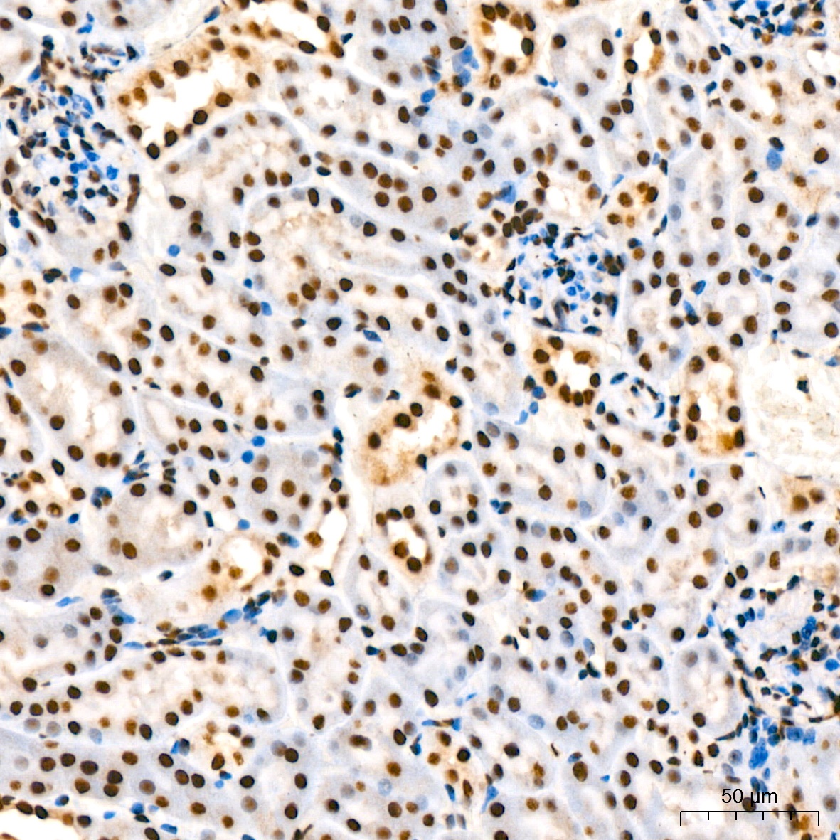
Immunohistochemistry analysis of paraffin-embedded Mouse kidney using Phospho-IκBα-S36 Rabbit mAb (CABP0999) at dilution of 1:200 (40x lens). High pressure antigen retrieval performed with 0.01M Citrate buffer (pH 6.0) prior to IHC staining.
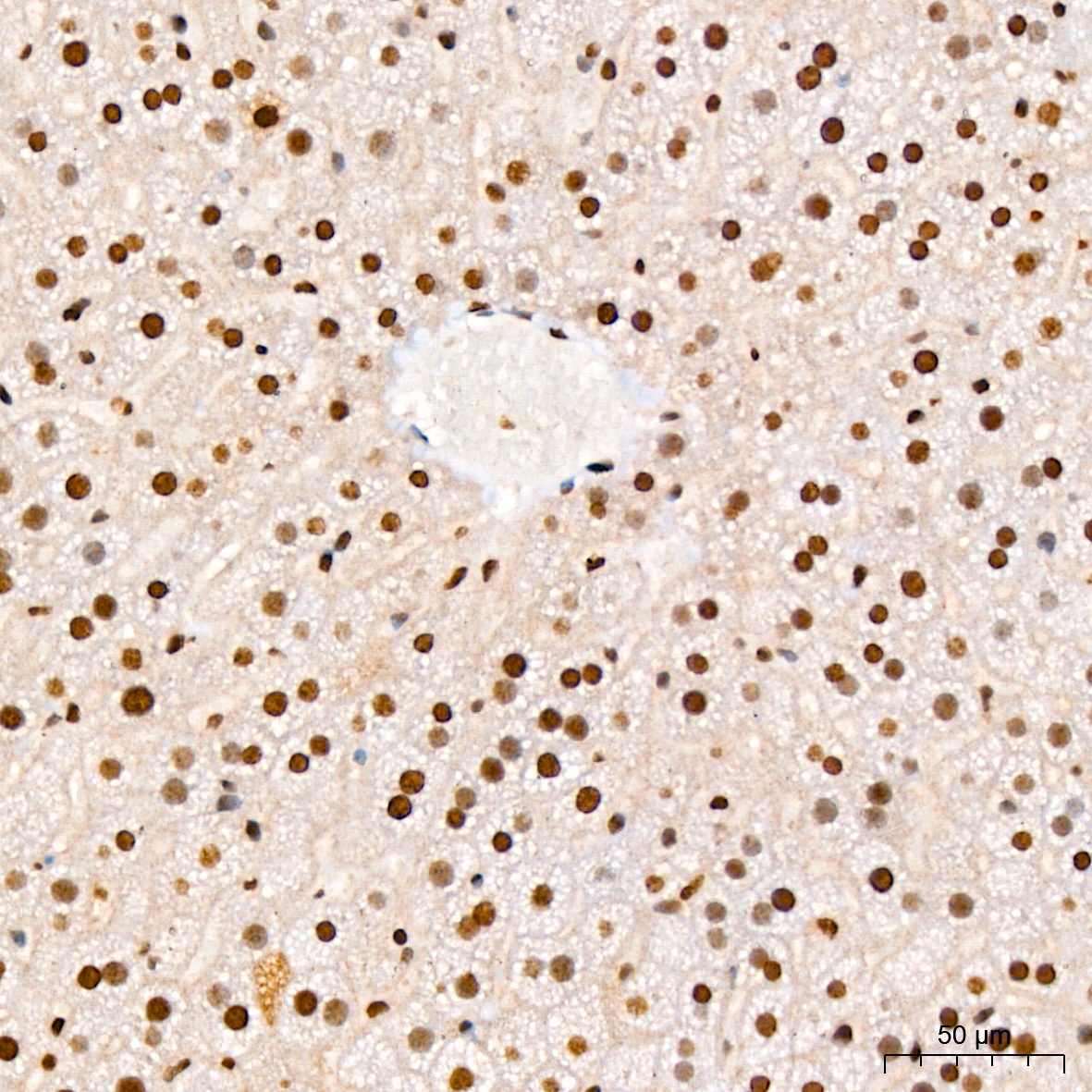
Immunohistochemistry analysis of paraffin-embedded Mouse liver using Phospho-IκBα-S36 Rabbit mAb (CABP0999) at dilution of 1:200 (40x lens). High pressure antigen retrieval performed with 0.01M Citrate buffer (pH 6.0) prior to IHC staining.