The Phospho-IkappaBalpha-S36 Antibody (CABP1069) is a high-quality antibody developed for reliable detection and analysis of target proteins. This antibody, produced in rabbits, exhibits high specificity for human samples and has been validated for use in Western blotting applications.IKBAlpha is a crucial component of the NF-κB pathway, which plays a central role in regulating inflammation, immune response, and cell survival. Phosphorylation of IKBAlpha at Serine 36 is known to trigger its degradation, leading to the activation of NF-κB transcription factors and the subsequent expression of inflammatory genes.
This antibody is validated for use in WB, ELISA applications and has demonstrated reactivity against Human, Mouse, Rat samples.
Product Name:
Phospho-IkappaBalpha-S36 Antibody
SKU:
CABP1069
Size:
20μL, 100μL
Reactivity:
Human, Mouse, Rat
Immunogen:
Synthetic peptide. This information is considered to be commercially sensitive.
Sequence:
LDSM K
Tested Applications:
WBELISA
Recommended Dilution:
WB
1:500 - 1:2000
ELISA
Recommended starting concentration is 1 μg/mL. Please optimize the concentration based on your specific assay requirements.
Synonyms:
IKBA, MAD-3, NFKBI, EDAID2, Phospho-IκBα-S36
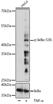
Positive Sample:
HeLa treated with TNF-α
Cellular Localization:
Cytoplasm, Nucleus.
Calculated MW:
36kDa
Observed MW:
45kDa
This gene encodes a member of the NF-kappa-B inhibitor family, which contain multiple ankrin repeat domains. The encoded protein interacts with REL dimers to inhibit NF-kappa-B/REL complexes which are involved in inflammatory responses. The encoded protein moves between the cytoplasm and the nucleus via a nuclear localization signal and CRM1-mediated nuclear export. Mutations in this gene have been found in ectodermal dysplasia anhidrotic with T-cell immunodeficiency autosomal dominant disease.
Purification Method
Affinity purification
Gene ID
4792
RRID
AB_2863941
Buffer Information
Store at -20℃. Avoid freeze / thaw cycles. Buffer: PBS containing 50% glycerol, preserved with proclin300 or sodium azide, pH 7.3.
Western blot analysis of lysates from HeLa cells, using Phospho-IκBα-S36 Rabbit pAb (CAB11397). HeLa cells were treated with TNF-α (20 ng/mL) at 37℃ for 30 minutes. Secondary antibody: HRP-conjugated Goat anti-Rabbit IgG (H+L) (CABS014) at 1:10000 dilution. Lysates/proteins: 25μg per lane. Blocking buffer: 3% BSA. Detection: ECL Basic Kit (AbGn00020). Exposure time: 30s.