The IRE1 Antibody (CAB17940) is a high-quality antibody developed for reliable detection and analysis of target proteins. This gene encodes the transmembrane protein kinase inositol-requiring enzyme 1. The encoded protein contains two functional catalytic domains, a serine/threonine-protein kinase domain and an endoribonuclease domain. This protein functions as a sensor of unfolded proteins in the endoplasmic reticulum (ER) and triggers an intracellular signaling pathway termed the unfolded protein response (UPR). The UPR is an ER stress response that is conserved from yeast to mammals and activates genes involved in degrading misfolded proteins, regulating protein synthesis and activating molecular chaperones. This protein specifically mediates the splicing and activation of the stress response transcription factor X-box binding protein 1.
This antibody is validated for use in WB, IHC-P, IF/ICC, ELISA applications and has demonstrated reactivity against Human, Mouse, Rat samples.
Product Name:
IRE1 Antibody
SKU:
CAB17940
Size:
100μL, 20μL
Reactivity:
Human, Mouse, Rat
Conjugate:
Unconjugated
Immunogen:
Recombinant protein (or fragment).This information is considered to be commercially sensitive.
Tested Applications:
WBIHC-PIF/ICCELISA
Recommended Dilution:
WB
1:500 - 1:5000
IF/ICC
1:50 - 1:200
IHC-P
1:50 - 1:200
ELISA
Recommended starting concentration is 1 μg/mL. Please optimize the concentration based on your specific assay requirements.
Synonyms:
IRE1, IRE1P, IRE1a, hIRE1p
Positive Sample:
293T, Mouse liver, Rat liver
Cellular Localization:
Endoplasmic Reticulum Membrane, Single-Pass Type I Membrane Protein.
Calculated MW:
110 kDa
Observed MW:
130 kDa
This gene encodes the transmembrane protein kinase inositol-requiring enzyme 1. The encoded protein contains two functional catalytic domains, a serine/threonine-protein kinase domain and an endoribonuclease domain. This protein functions as a sensor of unfolded proteins in the endoplasmic reticulum (ER) and triggers an intracellular signaling pathway termed the unfolded protein response (UPR). The UPR is an ER stress response that is conserved from yeast to mammals and activates genes involved in degrading misfolded proteins, regulating protein synthesis and activating molecular chaperones. This protein specifically mediates the splicing and activation of the stress response transcription factor X-box binding protein 1.
Purification Method
Affinity purification
Gene ID
2081
RRID
AB_2769358
Buffer Information
Store at -20℃. Avoid freeze / thaw cycles. Buffer: PBS with 0.09% Sodium azide,50% glycerol,pH7.3.
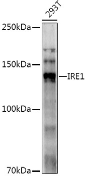
Western blot analysis of lysates from 293T cells, using IRE1 Rabbit pAb (CAB17940) at 1:1000 dilution. Secondary antibody: HRP-conjugated Goat anti-Rabbit IgG (H+L) (AS014) at 1:10000 dilution. Lysates/proteins: 25μg per lane. Blocking buffer: 3% nonfat dry milk in TBST. Detection: ECL Enhanced Kit (AbGn00021). Exposure time: 120s.
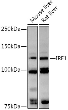
Western blot analysis of various lysates using IRE1 Rabbit pAb (CAB17940) at 1:1000 dilution. Secondary antibody: HRP-conjugated Goat anti-Rabbit IgG (H+L) (AS014) at 1:10000 dilution. Lysates/proteins: 25μg per lane. Blocking buffer: 3% nonfat dry milk in TBST. Detection: ECL Basic Kit (AbGn00020). Exposure time: 90s.
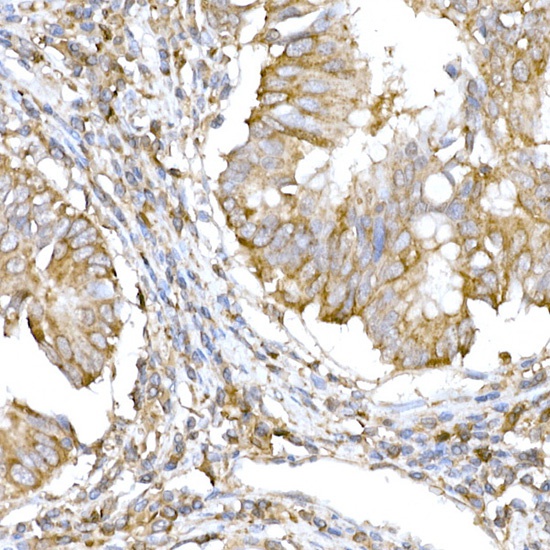
Immunohistochemistry analysis of paraffin-embedded Human colon carcinoma using IRE1 Rabbit pAb (CAB17940) at dilution of 1:100 (40x lens). High pressure antigen retrieval performed with 0.01M Citrate buffer (pH 6.0) prior to IHC staining.
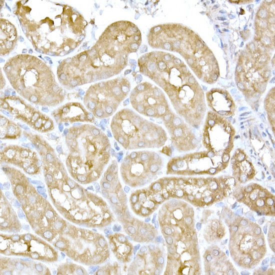
Immunohistochemistry analysis of paraffin-embedded Mouse kidney using IRE1 Rabbit pAb (CAB17940) at dilution of 1:100 (40x lens). High pressure antigen retrieval performed with 0.01M Citrate buffer (pH 6.0) prior to IHC staining.
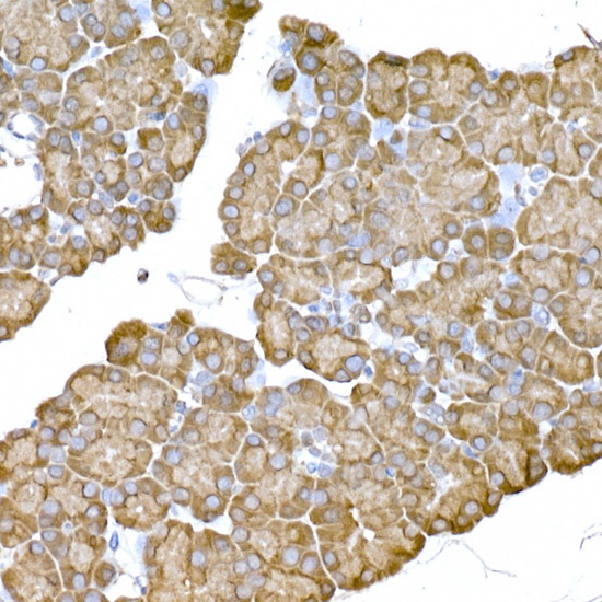
Immunohistochemistry analysis of paraffin-embedded Rat pancreas using IRE1 Rabbit pAb (CAB17940) at dilution of 1:100 (40x lens). High pressure antigen retrieval performed with 0.01M Citrate buffer (pH 6.0) prior to IHC staining.
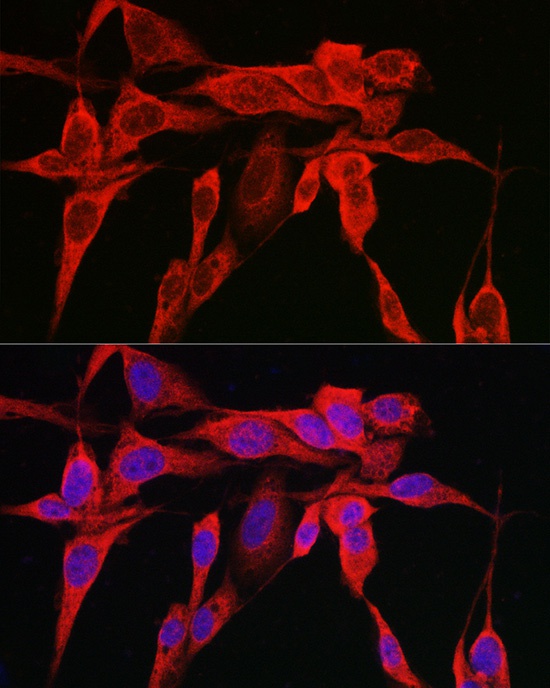
Immunofluorescence analysis of U2OS cells using IRE1 Rabbit pAb (CAB17940) at dilution of 1:100 (40x lens). Secondary antibody: Cy3-conjugated Goat anti-Rabbit IgG (H+L) (AS007) at 1:500 dilution. Blue: DAPI for nuclear staining.
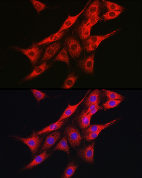
Immunofluorescence analysis of PC-12 cells using IRE1 Rabbit pAb (CAB17940) at dilution of 1:100 (40x lens). Secondary antibody: Cy3-conjugated Goat anti-Rabbit IgG (H+L) (AS007) at 1:500 dilution. Blue: DAPI for nuclear staining.