The Phospho-NMDAR1-S896/S897 Antibody (CABP0962) is a high-quality antibody developed for reliable detection and analysis of target proteins. This antibody, produced in rabbits, is highly specific for phosphorylated NMDAR1 at serine 896 and 897 residues, allowing for precise detection and analysis in various experimental settings. It is validated for use in techniques such as Western blotting, immunofluorescence, and immunoprecipitation.Phosphorylation of NMDAR1 at serine 896 and 897 plays a crucial role in regulating the activity of NMDA receptors, which are involved in synaptic plasticity, learning, and memory. Dysregulation of NMDAR1 phosphorylation has been implicated in neurological disorders such as Alzheimer's disease, schizophrenia, and epilepsy.
This antibody is validated for use in WB, ELISA applications and has demonstrated reactivity against Human, Mouse, Rat samples.
Product Name:
Phospho-NMDAR1-S896/S897 Antibody
SKU:
CABP0962
Size:
20μL, 100μL
Reactivity:
Human, Mouse, Rat
Conjugate:
Unconjugated
Immunogen:
Synthetic peptide. This information is considered to be commercially sensitive.
Sequence:
RRSS KD
Tested Applications:
WBELISA
Recommended Dilution:
WB
1:500 - 1:2000
ELISA
Recommended starting concentration is 1 μg/mL. Please optimize the concentration based on your specific assay requirements.
The protein encoded by this gene is a critical subunit of N-methyl-D-aspartate receptors, members of the glutamate receptor channel superfamily which are heteromeric protein complexes with multiple subunits arranged to form a ligand-gated ion channel. These subunits play a key role in the plasticity of synapses, which is believed to underlie memory and learning. Cell-specific factors are thought to control expression of different isoforms, possibly contributing to the functional diversity of the subunits. Alternatively spliced transcript variants have been described.
Purification Method
Affinity purification
Gene ID
2902
Buffer Information
Store at -20℃. Avoid freeze / thaw cycles. Buffer: PBS with 0.01% thimerosal,50% glycerol,pH7.3.
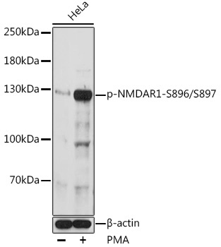
Western blot analysis of lysates from HeLa cells, using Phospho-NMDAR1-S896/S897 Rabbit pAb (CABP0962) at 1:1000 dilution. HeLa cells were treated with UV at room temperature for 30 minutes and PMA (200 nM) at 37℃ for 30 minutes after serum-starvation overnight. Secondary antibody: HRP-conjugated Goat anti-Rabbit IgG (H+L) (CABS014) at 1:10000 dilution. Lysates/proteins: 25μg per lane. Blocking buffer: 3% BSA. Detection: ECL Basic Kit (AbGn00020). Exposure time: 90s.