The PTBP2 Monoclonal Antibody (CAB9124) is a high-quality antibody developed for reliable detection and analysis of target proteins. This antibody, produced in rabbits, exhibits high specificity and sensitivity when used in applications such as Western blotting and immunohistochemistry. By binding to PTBP2, this antibody enables precise detection and analysis of the protein in various cells and tissues, making it indispensable for investigations in molecular biology and developmental biology.PTBP2, also known as polypyrimidine tract-binding protein 2, plays a crucial role in RNA processing and alternative splicing, thereby influencing the diversity of mRNA transcripts and protein isoforms generated in cells.
This antibody is validated for use in WB, IHC-P, IF/ICC, IP, ELISA, IF-P applications and has demonstrated reactivity against Human, Mouse, Rat samples.
Product Name:
PTBP2 Monoclonal Antibody
SKU:
CAB9124
Size:
20μL, 100μL
Reactivity:
Human, Mouse, Rat
Clone Number:
ARC1798
Conjugate:
Unconjugated
Immunogen:
Synthetic peptide. This information is considered to be commercially sensitive.
0.5μg-4μg antibody for 200μg-400μg extracts of whole cells
IF/ICC
1:200 - 1:2000
IF-P
1:200 - 1:2000
IHC-P
1:400 - 1:4000
ELISA
Recommended starting concentration is 1 μg/mL. Please optimize the concentration based on your specific assay requirements.
Synonyms:
nPTB, PTBLP, brPTB, PTBP2
Positive Sample:
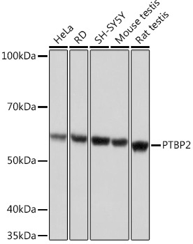
HeLa, RD, SH-SY5Y, Mouse testis, Rat testis
Cellular Localization:
Nucleus.
Calculated MW:
57kDa
Observed MW:
57kDa
The protein encoded by this gene binds to intronic polypyrimidine clusters in pre-mRNA molecules and is implicated in controlling the assembly of other splicing-regulatory proteins. This protein is very similar to the polypyrimidine tract binding protein (PTB) but most of its isoforms are expressed primarily in the brain. Alternative splicing results in multiple transcript variants.
Purification Method
Affinity purification
Gene ID
58155
RRID
AB_2863660
Buffer Information
Store at -20℃. Avoid freeze / thaw cycles. Buffer: PBS containing 50% glycerol and 0.05% BSA, preserved with proclin300 or sodium azide, pH 7.3.
Western blot analysis of various lysates using PTBP2 Rabbit mAb (CAB9124) at 1:1000 dilution. Secondary antibody: HRP-conjugated Goat anti-Rabbit IgG (H+L) (CABS014) at 1:10000 dilution. Lysates/proteins: 25μg per lane. Blocking buffer: 3% nonfat dry milk in TBST. Detection: ECL Basic Kit (AbGn00020). Exposure time: 1s.
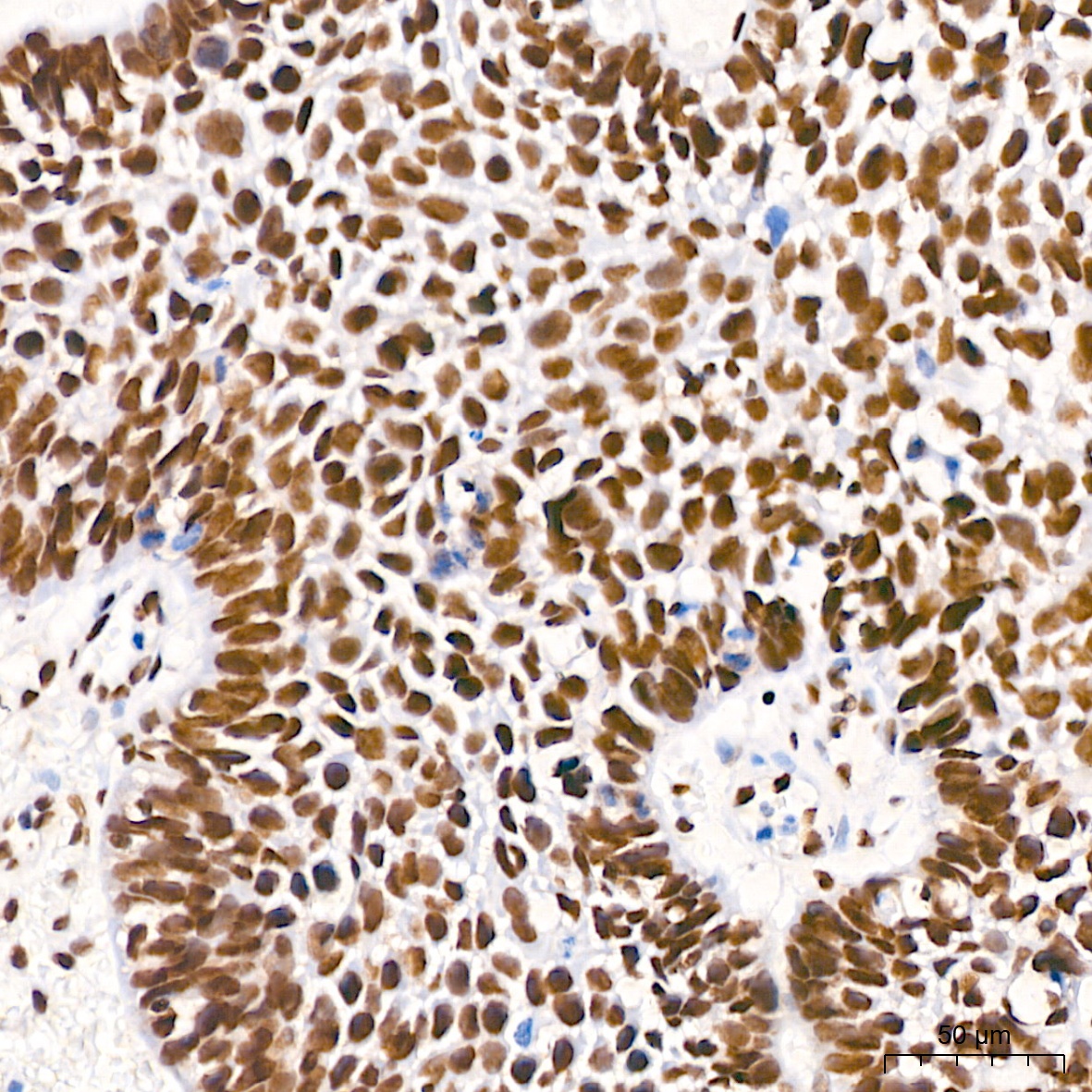
Immunohistochemistry analysis of paraffin-embedded Human cervix cancer tissue using PTBP2 Rabbit mAb (CAB9124) at a dilution of 1:800 (40x lens). High pressure antigen retrieval performed with 0.01M Tris-EDTA Buffer (pH 9.0) prior to IHC staining.
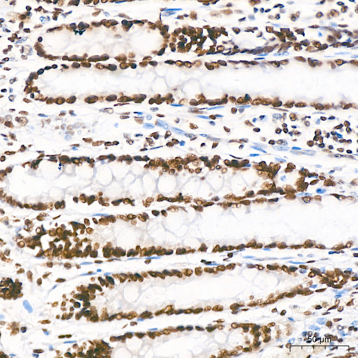
Immunohistochemistry analysis of paraffin-embedded Human colon tissue using PTBP2 Rabbit mAb (CAB9124) at a dilution of 1:800 (40x lens). High pressure antigen retrieval performed with 0.01M Tris-EDTA Buffer (pH 9.0) prior to IHC staining.
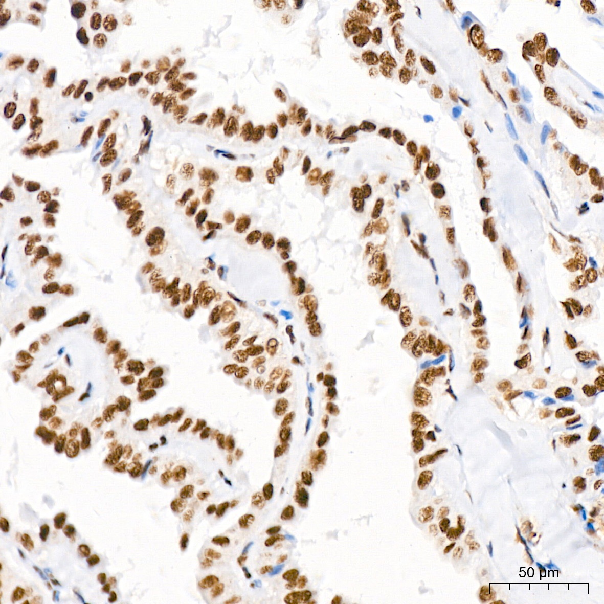
Immunohistochemistry analysis of paraffin-embedded Human thyroid cancer tissue using PTBP2 Rabbit mAb (CAB9124) at a dilution of 1:800 (40x lens). High pressure antigen retrieval performed with 0.01M Tris-EDTA Buffer (pH 9.0) prior to IHC staining.
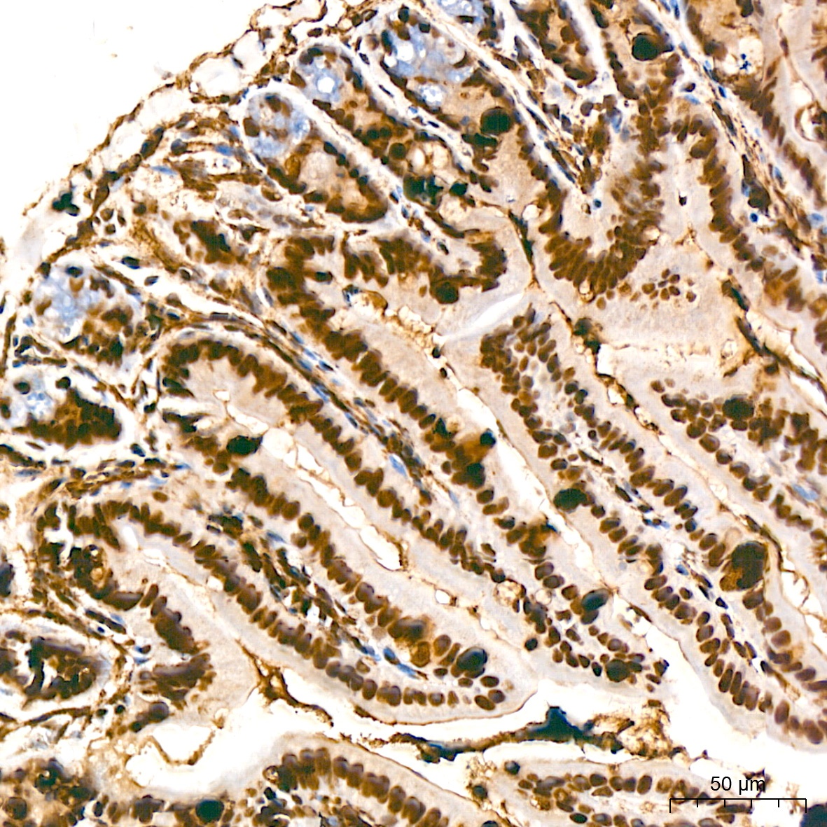
Immunohistochemistry analysis of paraffin-embedded Mouse intestin tissue using PTBP2 Rabbit mAb (CAB9124) at a dilution of 1:800 (40x lens). High pressure antigen retrieval performed with 0.01M Tris-EDTA Buffer (pH 9.0) prior to IHC staining.
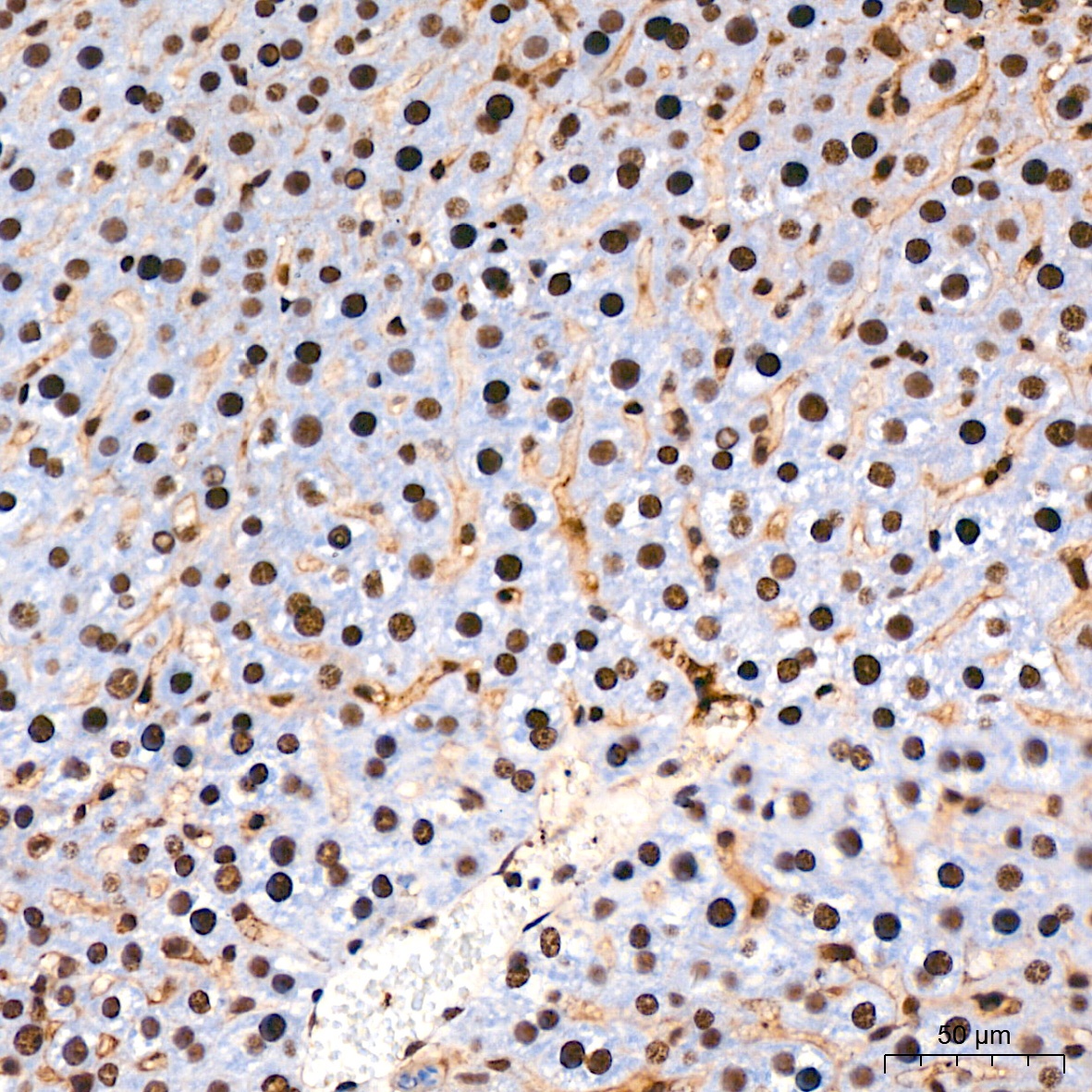
Immunohistochemistry analysis of paraffin-embedded Mouse liver tissue using PTBP2 Rabbit mAb (CAB9124) at a dilution of 1:800 (40x lens). High pressure antigen retrieval performed with 0.01M Tris-EDTA Buffer (pH 9.0) prior to IHC staining.
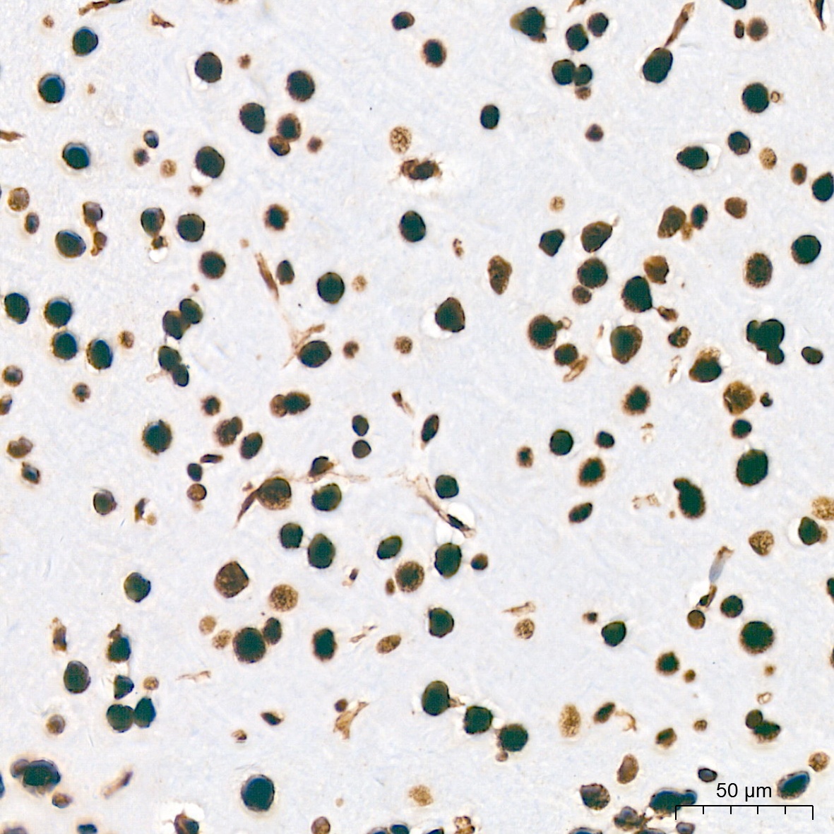
Immunohistochemistry analysis of paraffin-embedded Rat brain tissue using PTBP2 Rabbit mAb (CAB9124) at a dilution of 1:800 (40x lens). High pressure antigen retrieval performed with 0.01M Tris-EDTA Buffer (pH 9.0) prior to IHC staining.
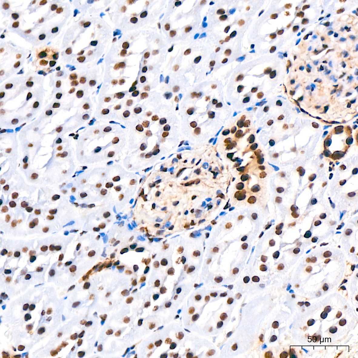
Immunohistochemistry analysis of paraffin-embedded Rat kidney tissue using PTBP2 Rabbit mAb (CAB9124) at a dilution of 1:800 (40x lens). High pressure antigen retrieval performed with 0.01M Tris-EDTA Buffer (pH 9.0) prior to IHC staining.
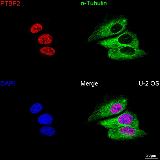
Confocal imaging of U-2 OS cells using PTBP2 Rabbit mAb (CAB9124, dilution 1:200) followed by a further incubation with Cy3 Goat Anti-Rabbit IgG (H+L) (CABS007, dilution 1:500) (Red). The cells were counterstained with α-Tubulin Mouse mAb (AC012, dilution 1:400) followed by incubation with ABflo® 488-conjugated Goat Anti-Mouse IgG (H+L) Ab (CABS076, dilution 1:500) (Green). DAPI was used for nuclear staining (Blue). Objective: 100x.
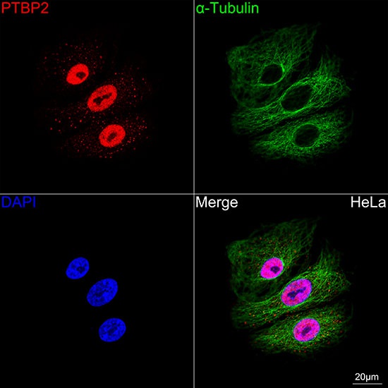
Confocal imaging of A549 cells using PTBP2 Rabbit mAb (CAB9124, dilution 1:200) followed by a further incubation with Cy3 Goat Anti-Rabbit IgG (H+L) (CABS007, dilution 1:500) (Red). The cells were counterstained with α-Tubulin Mouse mAb (AC012, dilution 1:400) followed by incubation with ABflo® 488-conjugated Goat Anti-Mouse IgG (H+L) Ab (CABS076, dilution 1:500) (Green). DAPI was used for nuclear staining (Blue). Objective: 100x.
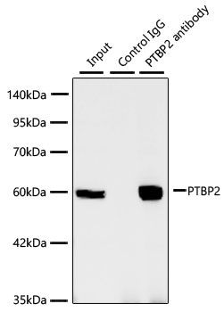
Immunoprecipitation of PTBP2 from 300 µg extracts of SH-SY5Y cells was performed using 3 µg of PTBP2 Rabbit mAb (CAB9124). Rabbit Control IgG (AC005) was used to precipitate the Control IgG sample. IP samples were eluted with 1X Laemmli Buffer. The Input lane represents 10% of the total input. Western blot analysis of immunoprecipitates was conducted using PTBP2 Rabbit mAb (CAB9124) at a dilution of 1:1000.