The QK1 Monoclonal Antibody (CAB0193) is a high-quality antibody developed for reliable detection and analysis of target proteins. This polyclonal antibody, produced in rabbits, exhibits high reactivity with human samples and is validated for use in various applications, including Western blotting and immunofluorescence.Qk1 is a RNA-binding protein that plays a critical role in post-transcriptional regulation of gene expression, especially in the nervous system. It is involved in processes such as RNA splicing, stability, and translation, making it a key player in neuronal development and function.
This antibody is validated for use in WB, IHC-P, IF/ICC, IP, ELISA applications and has demonstrated reactivity against Human, Mouse, Rat samples.
Product Name:
QK1 Monoclonal Antibody
SKU:
CAB0193
Size:
20μL, 100μL
Reactivity:
Human, Mouse, Rat
Clone Number:
ARC2500
Conjugate:
Unconjugated
Immunogen:
Synthetic peptide. This information is considered to be commercially sensitive.
0.5μg-4μg antibody for 200μg-400μg extracts of whole cells
ELISA
Recommended starting concentration is 1 μg/mL. Please optimize the concentration based on your specific assay requirements.
Synonyms:
QK, Hqk, QK1, QK3, hqkI, QKI
Positive Sample:
SKOV3, NIH/3T3, U-251MG, Neuro-2a, Mouse brain, Rat brain, Rat heart
Cellular Localization:
Cytoplasm, Nucleus.
Calculated MW:
38kDa
Observed MW:
38kDa/40kDa
The protein encoded by this gene is an RNA-binding protein that regulates pre-mRNA splicing, export of mRNAs from the nucleus, protein translation, and mRNA stability. The encoded protein is involved in myelinization and oligodendrocyte differentiation and may play a role in schizophrenia. Multiple transcript variants encoding different isoforms have been found for this gene.
Purification Method
Affinity purification
Gene ID
9444
Buffer Information
Store at -20℃. Avoid freeze / thaw cycles. Buffer: PBS containing 50% glycerol and 0.05% BSA, preserved with proclin300 or sodium azide, pH 7.3.
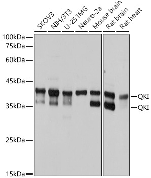
Western blot analysis of various lysates using QKI Rabbit mAb (CAB0193) at 1:1000 dilution. Secondary antibody: HRP-conjugated Goat anti-Rabbit IgG (H+L) (CABS014) at 1:10000 dilution. Lysates/proteins: 25μg per lane. Blocking buffer: 3% nonfat dry milk in TBST. Detection: ECL Basic Kit (AbGn00020). Exposure time: 1s.
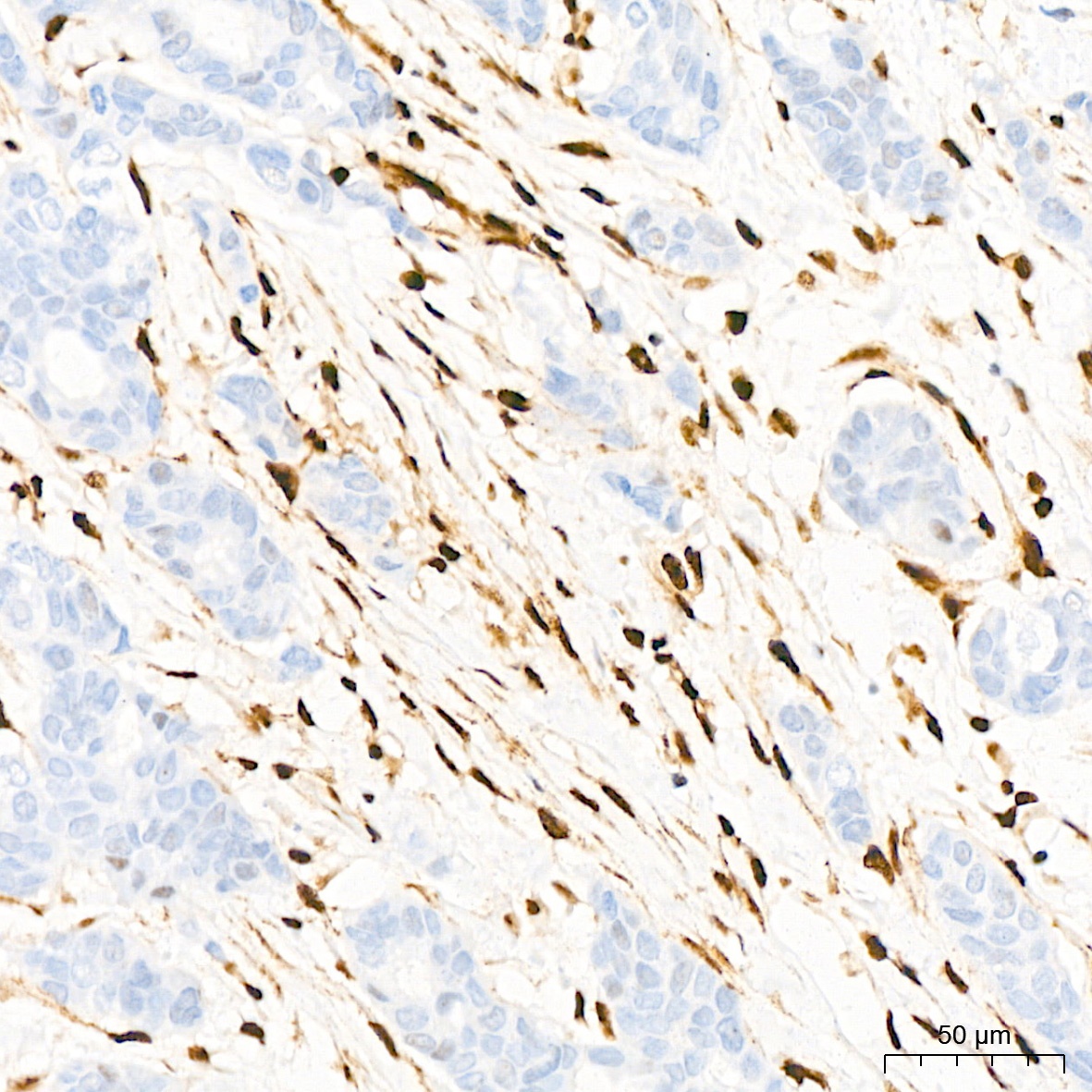
Immunohistochemistry analysis of paraffin-embedded Human breast cancer tissue using QKI Rabbit mAb (CAB0193) at a dilution of 1:200 (40x lens). High pressure antigen retrieval performed with 0.01M Citrate buffer (pH 6.0) prior to IHC staining.
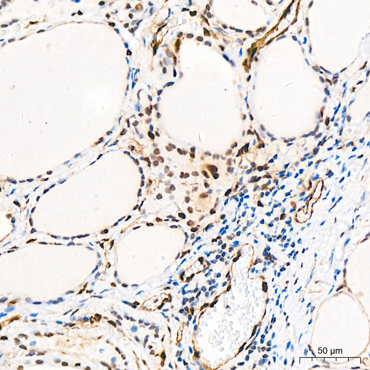
Immunohistochemistry analysis of paraffin-embedded Human thyroid tissue using QKI Rabbit mAb (CAB0193) at a dilution of 1:200 (40x lens). High pressure antigen retrieval performed with 0.01M Citrate buffer (pH 6.0) prior to IHC staining.
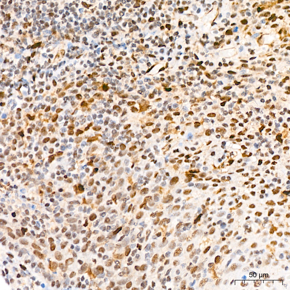
Immunohistochemistry analysis of paraffin-embedded Human tonsil tissue using QKI Rabbit mAb (CAB0193) at a dilution of 1:200 (40x lens). High pressure antigen retrieval performed with 0.01M Citrate buffer (pH 6.0) prior to IHC staining.
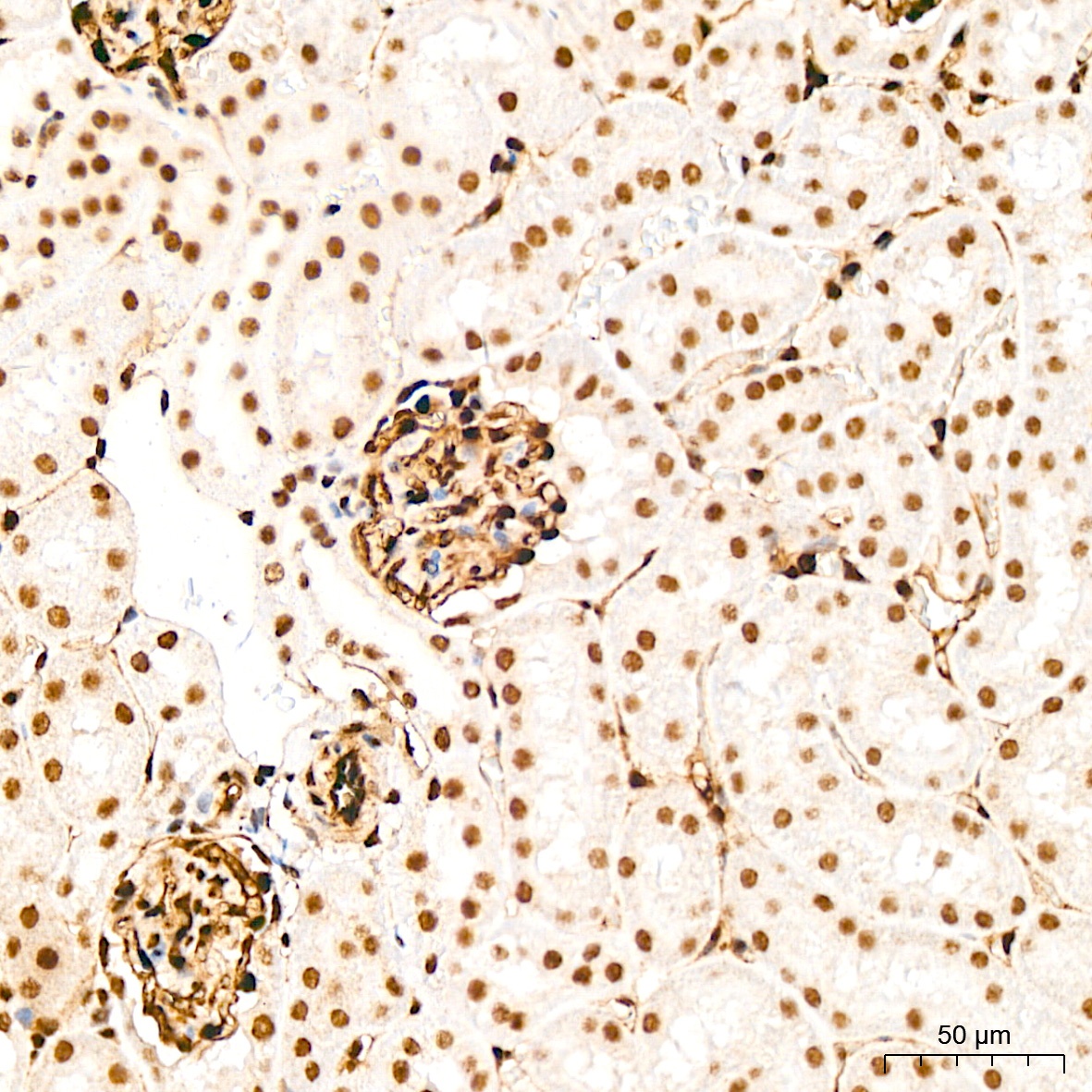
Immunohistochemistry analysis of paraffin-embedded Mouse kidney tissue using QKI Rabbit mAb (CAB0193) at a dilution of 1:200 (40x lens). High pressure antigen retrieval performed with 0.01M Citrate buffer (pH 6.0) prior to IHC staining.
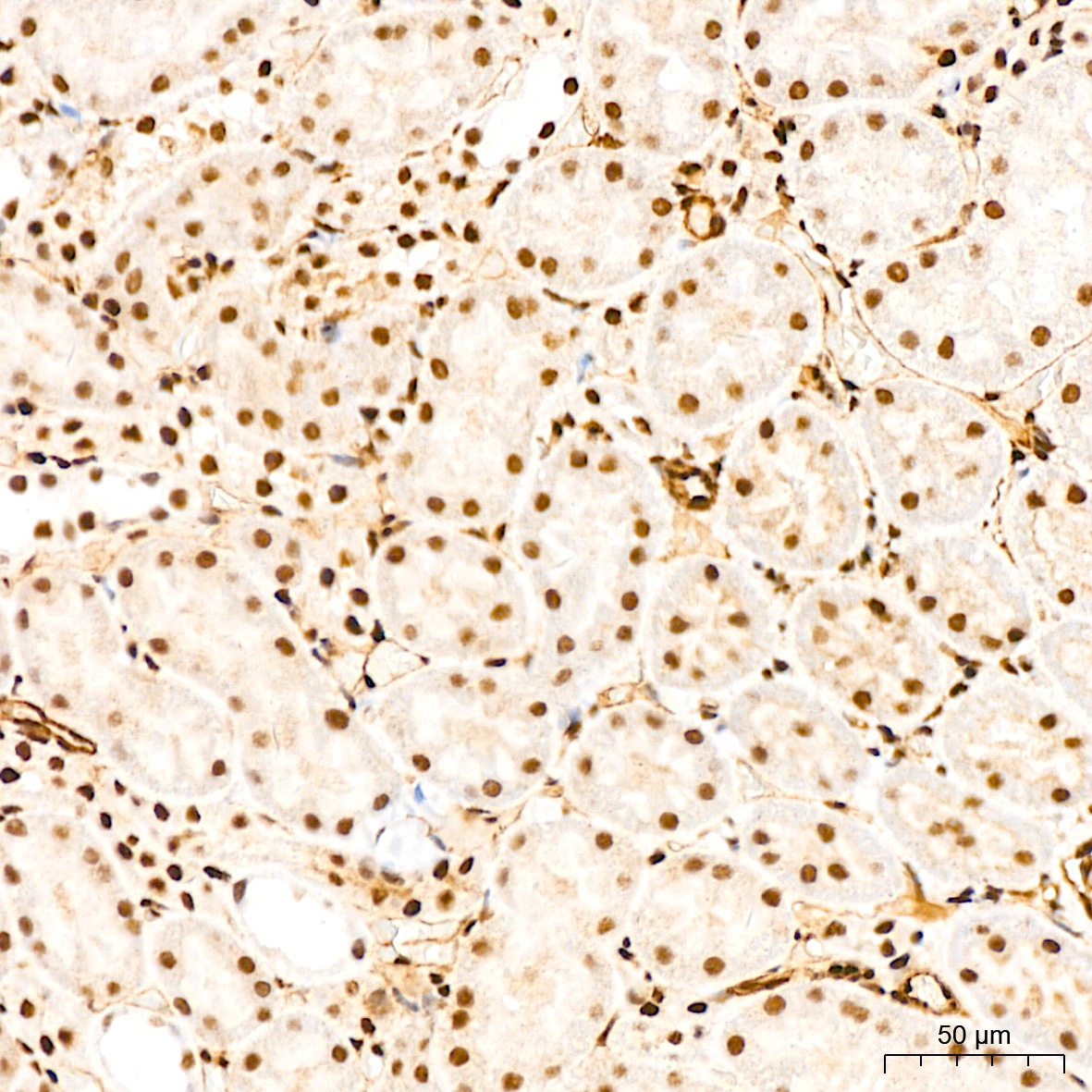
Immunohistochemistry analysis of paraffin-embedded Rat kidney tissue using QKI Rabbit mAb (CAB0193) at a dilution of 1:200 (40x lens). High pressure antigen retrieval performed with 0.01M Citrate buffer (pH 6.0) prior to IHC staining.
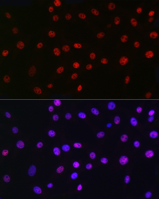
Immunofluorescence analysis of NIH/3T3 cells using QKI Rabbit mAb (CAB0193) at dilution of 1:100 (40x lens). Secondary antibody: Cy3-conjugated Goat anti-Rabbit IgG (H+L) (CABS007) at 1:500 dilution. Blue: DAPI for nuclear staining.