The Rad21 Antibody (CAB18850) is a high-quality antibody developed for reliable detection and analysis of target proteins. The protein encoded by this gene is highly similar to the gene product of Schizosaccharomyces pombe rad21, a gene involved in the repair of DNA double-strand breaks, as well as in chromatid cohesion during mitosis. This protein is a nuclear phospho-protein, which becomes hyperphosphorylated in cell cycle M phase. The highly regulated association of this protein with mitotic chromatin specifically at the centromere region suggests its role in sister chromatid cohesion in mitotic cells.
This antibody is validated for use in WB, IHC-P, IF/ICC, IP, ChIP, ELISA applications and has demonstrated reactivity against Human, Mouse, Rat samples.
Product Name:
Rad21 Antibody
SKU:
CAB18850
Size:
100μL, 20μL
Reactivity:
Human, Mouse, Rat
Immunogen:
Recombinant protein (or fragment).This information is considered to be commercially sensitive.
Tested Applications:
WBIHC-PIF/ICCIPChIPELISA
Recommended Dilution:
WB
1:500 - 1:1000
IHC-P
1:50 - 1:200
IF/ICC
1:50 - 1:200
IP
0.5μg-4μg antibody for 200μg-400μg extracts of whole cells
ELISA
Recommended starting concentration is 1 μg/mL. Please optimize the concentration based on your specific assay requirements.
Condensed Nuclear Chromosome, Cytosol, Nuclear Matrix, Nucleoplasm, Nucleus, Spindle Pole.
Calculated MW:
72kDa
Observed MW:
130kDa
The protein encoded by this gene is highly similar to the gene product of Schizosaccharomyces pombe rad21, a gene involved in the repair of DNA double-strand breaks, as well as in chromatid cohesion during mitosis. This protein is a nuclear phospho-protein, which becomes hyperphosphorylated in cell cycle M phase. The highly regulated association of this protein with mitotic chromatin specifically at the centromere region suggests its role in sister chromatid cohesion in mitotic cells.
Purification Method
Affinity purification
Gene ID
5885
RRID
AB_2862479
Buffer Information
Store at -20℃. Avoid freeze / thaw cycles. Buffer: PBS containing 50% glycerol, preserved with proclin300 or sodium azide, pH 7.3.
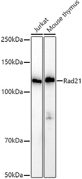
Western blot analysis of various lysates using Rad21 Rabbit pAb (CAB18850) at 1:1000 dilution. Secondary antibody: HRP-conjugated Goat anti-Rabbit IgG (H+L) (AS014) at 1:10000 dilution. Lysates/proteins: 25μg per lane. Blocking buffer: 3% nonfat dry milk in TBST. Detection: ECL Basic Kit (AbGn00020). Exposure time: 10s.
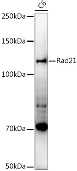
Western blot analysis of lysates from C6 cells, using Rad21 Rabbit pAb (CAB18850) at 1:1000 dilution. Secondary antibody: HRP-conjugated Goat anti-Rabbit IgG (H+L) (AS014) at 1:10000 dilution. Lysates/proteins: 25μg per lane. Blocking buffer: 3% nonfat dry milk in TBST. Detection: ECL Enhanced Kit (AbGn00021). Exposure time: 90s.
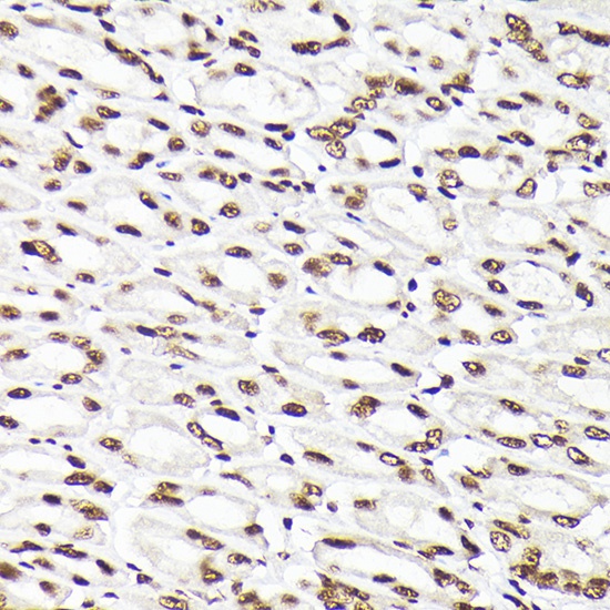
Immunohistochemistry analysis of paraffin-embedded Mouse stomach using Rad21 Rabbit pAb (CAB18850) at dilution of 1:200 (40x lens). High pressure antigen retrieval performed with 0.01M Citrate buffer (pH 6.0) prior to IHC staining.
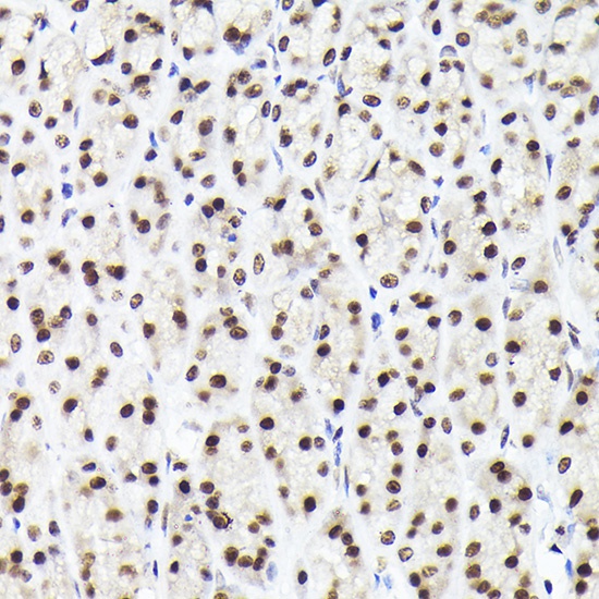
Immunohistochemistry analysis of paraffin-embedded Rat stomach using Rad21 Rabbit pAb (CAB18850) at dilution of 1:200 (40x lens). High pressure antigen retrieval performed with 0.01M Citrate buffer (pH 6.0) prior to IHC staining.
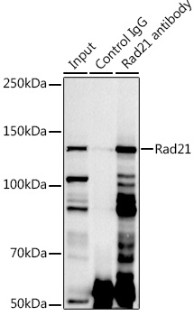
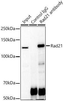
Immunoprecipitation analysis of 300 μg extracts of Jurkat cells using 3 μg Rad21 antibody (CAB18850). Western blot was performed from the immunoprecipitate using Rad21 antibody (CAB18850) at a dilution of 1:1000.
Immunoprecipitation analysis of 300 μg extracts of Jurkat cells using 3 μg Rad21 antibody (CAB18850). Western blot was performed from the immunoprecipitate using Rad21 antibody (CAB18850) at a dilution of 1:1000.
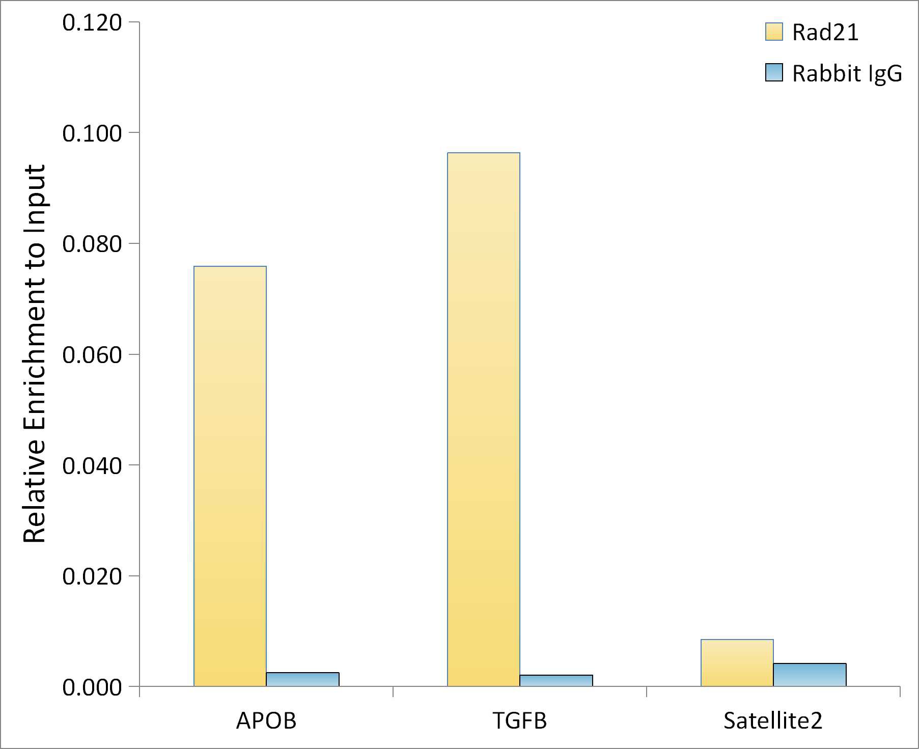
Chromatin immunoprecipitation analysis of extracts of A-549 cells, using Rad21 antibody (CAB18850) and rabbit IgG.The amount of immunoprecipitated DNA was checked by quantitative PCR. Histogram was constructed by the ratios of the immunoprecipitated DNA to the input.