The S100A6 Monoclonal Antibody (CAB3461) is a high-quality antibody developed for reliable detection and analysis of target proteins. This antibody, generated in rabbits, exhibits high specificity and sensitivity when detecting S100A6 in human samples, making it suitable for Western blot applications. By targeting the S100A6 protein, this antibody enables precise detection and analysis in a variety of cell types, making it a versatile tool for studies in cancer biology, neurology, and inflammation research.S100A6 has been implicated in various cellular processes, including cell cycle regulation, apoptosis, and inflammation, making it a key player in the development and progression of diseases such as cancer and neurodegenerative disorders.
This antibody is validated for use in WB, IHC-P, IF/ICC, ELISA applications and has demonstrated reactivity against Human, Mouse, Rat samples.
Product Name:
S100A6 Monoclonal Antibody
SKU:
CAB3461
Size:
20μL, 100μL
Reactivity:
Human, Mouse, Rat
Clone Number:
ARC2005
Conjugate:
Unconjugated
Immunogen:
Recombinant protein (or fragment).This information is considered to be commercially sensitive.
The protein encoded by this gene is a member of the S100 family of proteins containing 2 EF-hand calcium-binding motifs. S100 proteins are localized in the cytoplasm and/or nucleus of a wide range of cells, and involved in the regulation of a number of cellular processes such as cell cycle progression and differentiation. S100 genes include at least 13 members which are located as a cluster on chromosome 1q21. This protein may function in stimulation of Ca2+-dependent insulin release, stimulation of prolactin secretion, and exocytosis. Chromosomal rearrangements and altered expression of this gene have been implicated in melanoma.
Purification Method
Affinity purification
Gene ID
6277
RRID
AB_2863063
Buffer Information
Store at -20℃. Avoid freeze / thaw cycles. Buffer: PBS containing 50% glycerol and 0.05% BSA, preserved with proclin300 or sodium azide, pH 7.3.
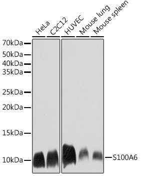
Western blot analysis of various lysates using S100A6 Rabbit mAb (CAB3461) at 1:1000 dilution. Secondary antibody: HRP-conjugated Goat anti-Rabbit IgG (H+L) (CABS014) at 1:10000 dilution. Lysates/proteins: 25μg per lane. Blocking buffer: 3% nonfat dry milk in TBST. Detection: ECL Basic Kit (AbGn00020). Exposure time: 30s.
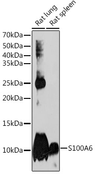
Western blot analysis of various lysates using S100A6 Rabbit mAb (CAB3461) at 1:1000 dilution. Secondary antibody: HRP-conjugated Goat anti-Rabbit IgG (H+L) (CABS014) at 1:10000 dilution. Lysates/proteins: 25μg per lane. Blocking buffer: 3% nonfat dry milk in TBST. Detection: ECL Basic Kit (AbGn00020). Exposure time: 3min.
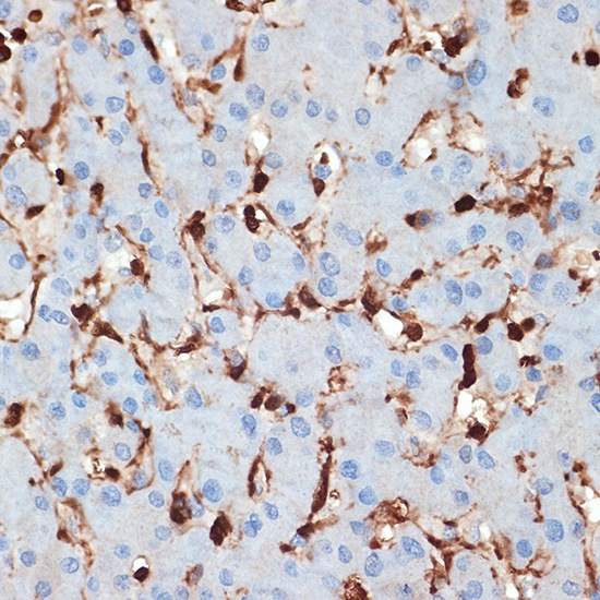
Immunohistochemistry analysis of paraffin-embedded Human liver using S100A6 Rabbit mAb (CAB3461) at dilution of 1:100 (40x lens). Microwave antigen retrieval performed with 0.01M Tris/EDTA Buffer (pH 9.0) prior to IHC staining.
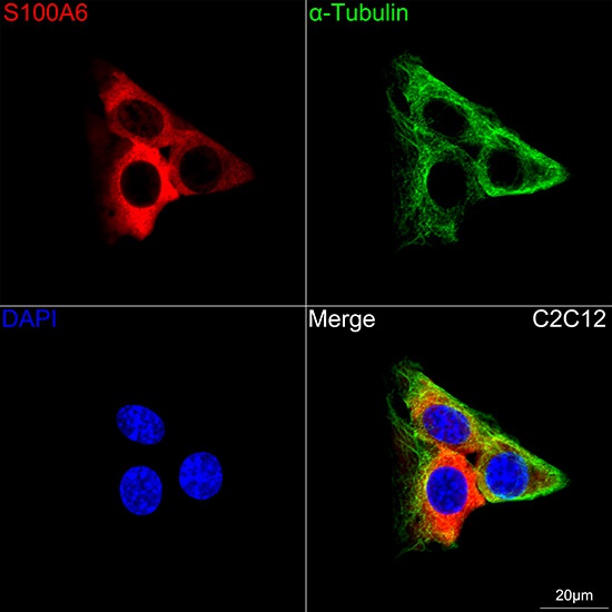
Confocal imaging of C2C12 cells using S100A6 Rabbit mAb (CAB3461, dilution 1:400) followed by a further incubation with Cy3 Goat Anti-Rabbit IgG (H+L) (CABS007, dilution 1:500) (Red). The cells were counterstained with α-Tubulin Mouse mAb (AC012, dilution 1:400) followed by incubation with ABflo® 488-conjugated Goat Anti-Mouse IgG (H+L) Ab (CABS076, dilution 1:500) (Green). DAPI was used for nuclear staining (Blue). Objective: 100x.