The Slit2 Monoclonal Antibody (CAB3467) is a high-quality antibody developed for reliable detection and analysis of target proteins. This polyclonal antibody, raised in rabbits, is highly specific to Slit2 and is validated for use in a variety of applications, including immunohistochemistry and immunocytochemistry.Slit2 is a secreted protein that plays a crucial role in axon guidance, tissue morphogenesis, and angiogenesis. It is known to interact with several receptors, including Robo1 and Robo2, to mediate cellular responses.
This antibody is validated for use in WB, IHC-P, ELISA applications and has demonstrated reactivity against Human, Mouse, Rat samples.
Product Name:
Slit2 Monoclonal Antibody
SKU:
CAB3467
Size:
20μL, 100μL
Reactivity:
Human, Mouse, Rat
Clone Number:
ARC2010
Conjugate:
Unconjugated
Immunogen:
Synthetic peptide. This information is considered to be commercially sensitive.
Recommended starting concentration is 1 μg/mL. Please optimize the concentration based on your specific assay requirements.
Synonyms:
SLIL3, Slit-2, Slit2
Positive Sample:
293T, PC-3
Cellular Localization:
Secreted.
Calculated MW:
170kDa
Observed MW:
200kDa
This gene encodes a member of the slit family of secreted glycoproteins, which are ligands for the Robo family of immunoglobulin receptors. Slit proteins play highly conserved roles in axon guidance and neuronal migration and may also have functions during other cell migration processes including leukocyte migration. Members of the slit family are characterized by an N-terminal signal peptide, four leucine-rich repeats, nine epidermal growth factor repeats, and a C-terminal cysteine knot. Proteolytic processing of this protein gives rise to an N-terminal fragment that contains the four leucine-rich repeats and five epidermal growth factor repeats and a C-terminal fragment that contains four epidermal growth factor repeats and the cysteine knot. Both full length and cleaved proteins are secreted extracellularly and can function in axon repulsion as well as other specific processes. Alternative splicing results in multiple transcript variants.
Purification Method
Affinity purification
Gene ID
9353
RRID
AB_2863066
Buffer Information
Store at -20℃. Avoid freeze / thaw cycles. Buffer: PBS containing 50% glycerol and 0.05% BSA, preserved with proclin300 or sodium azide, pH 7.3.
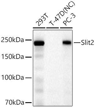
Western blot analysis of various lysates using Slit2 Rabbit mAb (CAB3467) at 1:1000 dilution incubated overnight at 4℃. Secondary antibody: HRP-conjugated Goat anti-Rabbit IgG (H+L) (CABS014) at 1:10000 dilution. Lysates/proteins: 25 μg per lane. Blocking buffer: 3% nonfat dry milk in TBST. Detection: ECL Basic Kit (AbGn00020). Negative control (NC): T-47D. Exposure time: 45 s.
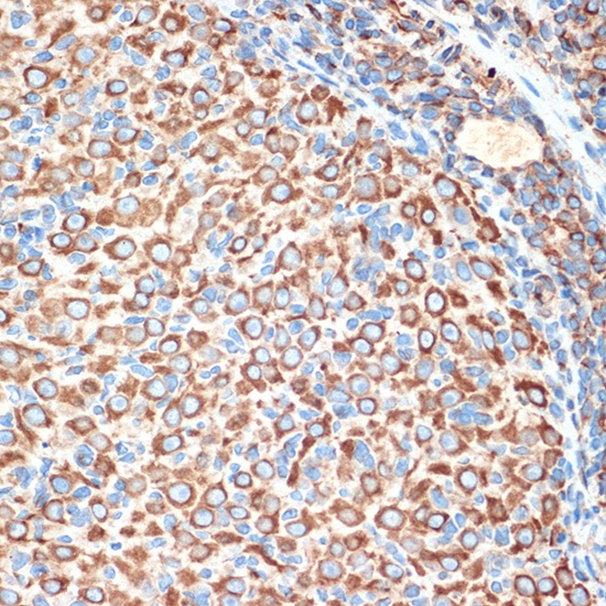
Immunohistochemistry analysis of paraffin-embedded Rat ovary using Slit2 Rabbit mAb (CAB3467) at dilution of 1:100 (40x lens). Microwave antigen retrieval performed with 0.01M Tris/EDTA Buffer (pH 9.0) prior to IHC staining.