The TBR1 Monoclonal Antibody (CAB19550) is a high-quality antibody developed for reliable detection and analysis of target proteins. This antibody, generated in rabbits, exhibits high specificity for human samples and has been validated for use in Western blot applications. By binding to the TBR1 protein, this antibody enables accurate detection and analysis in a variety of cell types, making it ideal for investigations in neurobiology and developmental disorders.
This antibody is validated for use in WB, ELISA, IF-P applications and has demonstrated reactivity against Mouse samples.
Product Name:
TBR1 Monoclonal Antibody
SKU:
CAB19550
Size:
20μL, 100μL
Reactivity:
Mouse
Clone Number:
ARC2198
Conjugate:
Unconjugated
Immunogen:
Synthetic peptide. This information is considered to be commercially sensitive.
Recommended starting concentration is 1 μg/mL. Please optimize the concentration based on your specific assay requirements.
Synonyms:
AUTS5, IDDAS, TBR-1, TES-56, TBR1
Positive Sample:
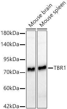
Mouse brain, Mouse spleen
Cellular Localization:
Nucleus.
Calculated MW:
74kDa
Observed MW:
74kDa
This gene is a member of a conserved family of genes that share a common DNA-binding domain, the T-box. T-box genes encode transcription factors involved in the regulation of numerous developmental processes. In mouse, the ortholog of this gene is expressed in the cerebral cortex, hippocampus, amygdala and olfactory bulb and is thought to play an important role in neuronal migration and axonal projection. In mouse, the C-terminal region of this protein was found to be necessary and sufficient for association with the guanylate kinase domain of calcium/calmodulin-dependent serine protein kinase.
Purification Method
Affinity purification
Gene ID
10716
Buffer Information
Store at -20℃. Avoid freeze / thaw cycles. Buffer: PBS containing 50% glycerol and 0.05% BSA, preserved with proclin300 or sodium azide, pH 7.3.
Western blot analysis of various lysates using TBR1 Rabbit mAb (CAB19550) at 1:1000 dilution incubated overnight at 4℃. Secondary antibody: HRP-conjugated Goat anti-Rabbit IgG (H+L) (CABS014) at 1:10000 dilution. Lysates/proteins: 25 μg per lane. Blocking buffer: 3% nonfat dry milk in TBST. Detection: ECL Basic Kit (AbGn00020). Exposure time: 45s.
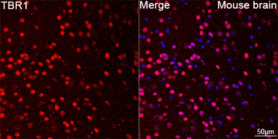
Confocal imaging of paraffin-embedded Mouse brain using TBR1 Rabbit mAb (CAB19550, dilution 1:200) followed by a further incubation with Cy3 Goat Anti-Rabbit IgG (H+L) (CABS007, dilution 1:500) (Red). DAPI was used for nuclear staining (Blue). Objective: 40x. Perform microwave antigen retrieval with 0.01 M citrate buffer (pH 6.0) prior to IF staining.