The TCF1/TCF7 Monoclonal Antibody (CAB20835) is a high-quality antibody developed for reliable detection and analysis of target proteins. This gene encodes a member of the T-cell factor/lymphoid enhancer-binding factor family of high mobility group (HMG) box transcriptional activators. This gene is expressed predominantly in T-cells and plays a critical role in natural killer cell and innate lymphoid cell development. The encoded protein forms a complex with beta-catenin and activates transcription through a Wnt/beta-catenin signaling pathway. Mice with a knockout of this gene are viable and fertile, but display a block in T-lymphocyte differentiation. Alternative splicing results in multiple transcript variants. Naturally-occurring isoforms lacking the N-terminal beta-catenin interaction domain may act as dominant negative regulators of Wnt signaling.
This antibody is validated for use in WB, ELISA applications and has demonstrated reactivity against Human, Mouse, Rat samples.
Product Name:
TCF1/TCF7 Monoclonal Antibody
SKU:
CAB20835
Size:
100μL, 20μL
Reactivity:
Human, Mouse, Rat
Clone Number:
ARC51870
Conjugate:
Unconjugated
Immunogen:
Synthetic peptide. This information is considered to be commercially sensitive.
Tested Applications:
WBELISA
Recommended Dilution:
WB
1:1000 - 1:2000
ELISA
Recommended starting concentration is 1 μg/mL. Please optimize the concentration based on your specific assay requirements.
Synonyms:
TCF-1, TCF1/TCF7
Positive Sample:
Jurkat, MOLT-4, Mouse thymus
Cellular Localization:
Nucleus.
Calculated MW:
42kDa
Observed MW:
28-50kDa
This gene encodes a member of the T-cell factor/lymphoid enhancer-binding factor family of high mobility group (HMG) box transcriptional activators. This gene is expressed predominantly in T-cells and plays a critical role in natural killer cell and innate lymphoid cell development. The encoded protein forms a complex with beta-catenin and activates transcription through a Wnt/beta-catenin signaling pathway. Mice with a knockout of this gene are viable and fertile, but display a block in T-lymphocyte differentiation. Alternative splicing results in multiple transcript variants. Naturally-occurring isoforms lacking the N-terminal beta-catenin interaction domain may act as dominant negative regulators of Wnt signaling.
Purification Method
Affinity purification
Gene ID
6932
Buffer Information
Store at -20℃. Avoid freeze / thaw cycles. Buffer: PBS containing 50% glycerol and 0.05% BSA, preserved with proclin300 or sodium azide, pH 7.3.
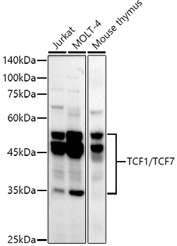
Western blot analysis of various lysates using TCF1/TCF7 Rabbit mAb (CAB20835) at1:1000 dilution. Secondary antibody: HRP-conjugated Goat anti-Rabbit IgG (H+L) (AS014) at1:10000 dilution. Lysates/proteins: 25μg per lane. Blocking buffer: 3% nonfat dry milk in TBST. Detection: ECL Basic Kit (AbGn00020). Exposure time: 180s.
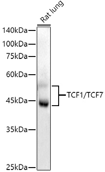
Western blot analysis of lysates from Rat lung, using TCF1/TCF7 Rabbit mAb (CAB20835) at1:1000 dilution. Secondary antibody: HRP-conjugated Goat anti-Rabbit IgG (H+L) (AS014) at1:10000 dilution. Lysates/proteins: 25μg per lane. Blocking buffer: 3% nonfat dry milk in TBST. Detection: ECL Enhanced Kit (AbGn00021). Exposure time: 90s.
. Perform high pressure antigen retrieval with 10 mM citrate buffer pH 6. 0 before commencing with IHC staining protocol.")
. Perform high pressure antigen retrieval with 10 mM citrate buffer pH 6. 0 before commencing with IHC staining protocol.")
. Perform high pressure antigen retrieval with 10 mM citrate buffer pH 6. 0 before commencing with IHC staining protocol.")
 at 1:10000 dilution. Lysates/proteins: 25ug per lane. Blocking buffer: 3% nonfat dry milk in TBST. Detection: ECL Enhanced Kit. Exposure time: 90s.")
. Perform high pressure antigen retrieval with 10 mM citrate buffer pH 6. 0 before commencing with IHC staining protocol.")
 at 1:10000 dilution. Lysates/proteins: 25ug per lane. Blocking buffer: 3% nonfat dry milk in TBST. Detection: ECL Basic Kit. Exposure time: 180s.")
. Perform high pressure antigen retrieval with 10 mM citrate buffer pH 6. 0 before commencing with IHC staining protocol.")
. Perform high pressure antigen retrieval with 10 mM citrate buffer pH 6. 0 before commencing with IHC staining protocol.")
 at 1:10000 dilution. Lysates/proteins: 25ug per lane. Blocking buffer: 3% nonfat dry milk in TBST. Detection: ECL Enhanced Kit. Exposure time: 90s.")
. Perform high pressure antigen retrieval with 10 mM citrate buffer pH 6. 0 before commencing with IHC staining protocol.")
 at 1:10000 dilution. Lysates/proteins: 25ug per lane. Blocking buffer: 3% nonfat dry milk in TBST. Detection: ECL Basic Kit. Exposure time: 180s.")


")
")
