The TNFRSF17 Antibody (CAB10707) is a high-quality antibody developed for reliable detection and analysis of target proteins. This polyclonal antibody, generated in rabbits, exhibits strong reactivity with human samples and is validated for use in Western blot applications.TNFRSF17, a member of the tumor necrosis factor receptor superfamily, plays a pivotal role in B cell survival and differentiation, making it a key target for investigating diseases such as multiple myeloma and other B cell-related disorders.
This antibody is validated for use in WB, IF/ICC, ELISA applications and has demonstrated reactivity against Human, Mouse, Rat samples.
Product Name:
TNFRSF17 Antibody
SKU:
CAB10707
Size:
20μL, 100μL
Reactivity:
Human, Mouse, Rat
Conjugate:
Unconjugated
Immunogen:
Synthetic peptide. This information is considered to be commercially sensitive.
Recommended starting concentration is 1 μg/mL. Please optimize the concentration based on your specific assay requirements.
Synonyms:
BCM, BCMA, CD269, TNFRSF13A, TNFRSF17
Positive Sample:
SP2/0, Raji, Rat spleen
Cellular Localization:
Cell Membrane, Endomembrane System, Single-Pass Type Iii Membrane Protein.
Calculated MW:
20kDa
Observed MW:
15-35kDa
The protein encoded by this gene is a member of the TNF-receptor superfamily. This receptor is preferentially expressed in mature B lymphocytes, and may be important for B cell development and autoimmune response. This receptor has been shown to specifically bind to the tumor necrosis factor (ligand) superfamily, member 13b (TNFSF13B/TALL-1/BAFF), and to lead to NF-kappaB and MAPK8/JNK activation. This receptor also binds to various TRAF family members, and thus may transduce signals for cell survival and proliferation.
Purification Method
Affinity purification
Gene ID
608
RRID
AB_2772656
Buffer Information
Store at -20℃. Avoid freeze / thaw cycles. Buffer: PBS containing 50% glycerol, preserved with proclin300 or sodium azide, pH 7.3.
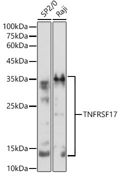
Western blot analysis of various lysates using TNFRSF17 Rabbit pAb (CAB10707) at 1:500 dilution. Secondary antibody: HRP-conjugated Goat anti-Rabbit IgG (H+L) (CABS014) at 1:10000 dilution. Lysates/proteins: 25μg per lane. Blocking buffer: 3% nonfat dry milk in TBST. Detection: ECL Basic Kit (AbGn00020). Exposure time: 180s.
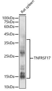
Western blot analysis of lysates from Rat spleen, using TNFRSF17 Rabbit pAb (CAB10707) at 1:500 dilution. Secondary antibody: HRP-conjugated Goat anti-Rabbit IgG (H+L) (CABS014) at 1:10000 dilution. Lysates/proteins: 25μg per lane. Blocking buffer: 3% nonfat dry milk in TBST. Detection: ECL Enhanced Kit (AbGn00021). Exposure time: 180s.
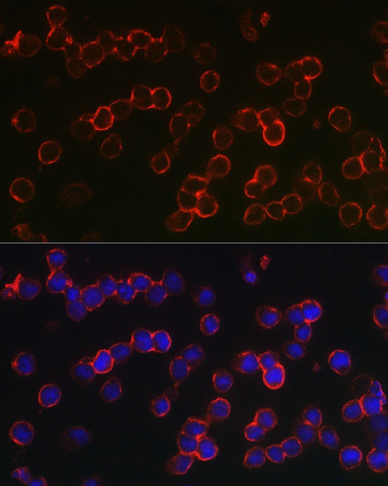
Immunofluorescence analysis of U-937 cells using TNFRSF17 Rabbit pAb (CAB10707) at dilution of 1:100 (40x lens). Secondary antibody: Cy3-conjugated Goat anti-Rabbit IgG (H+L) (CABS007) at 1:500 dilution. Blue: DAPI for nuclear staining.