The Torsin A Monoclonal Antibody (CAB9579) is a high-quality antibody developed for reliable detection and analysis of target proteins. This polyclonal antibody, produced in rabbits, is highly specific and reactive with human samples, making it an excellent choice for Western blot and immunofluorescence applications.Torsina is a key player in the pathogenesis of DYT1 dystonia, a movement disorder characterized by involuntary muscle contractions. By targeting Torsina with this antibody, researchers can gain insights into the molecular mechanisms underlying DYT1 dystonia and potentially identify new therapeutic targets for the treatment of this debilitating condition.
This antibody is validated for use in WB, IF/ICC, ELISA applications and has demonstrated reactivity against Human, Mouse, Rat samples.
Product Name:
Torsin A Monoclonal Antibody
SKU:
CAB9579
Size:
20μL, 100μL
Reactivity:
Human, Mouse, Rat
Clone Number:
ARC1645
Conjugate:
Unconjugated
Immunogen:
Synthetic peptide. This information is considered to be commercially sensitive.
The protein encoded by this gene is a member of the AAA family of adenosine triphosphatases (ATPases), is related to the Clp protease/heat shock family and is expressed prominently in the substantia nigra pars compacta. Mutations in this gene result in the autosomal dominant disorder, torsion dystonia 1.
Purification Method
Affinity purification
Gene ID
1861
Buffer Information
Store at -20℃. Avoid freeze / thaw cycles. Buffer: PBS containing 50% glycerol and 0.05% BSA, preserved with proclin300 or sodium azide, pH 7.3.
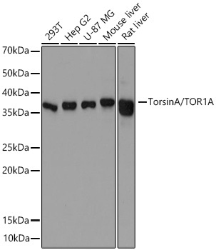
Western blot analysis of various lysates using TorsinA/TOR1A Rabbit mAb (CAB9579) at 1:500 dilution incubated at room temperature for 1.5 hours. Secondary antibody: HRP-conjugated Goat anti-Rabbit IgG (H+L) (CABS014) at 1:10000 dilution. Lysates/proteins: 25 μg per lane. Blocking buffer: 3% nonfat dry milk in TBST. Detection: ECL Basic Kit (AbGn00020). Exposure time: 60s.
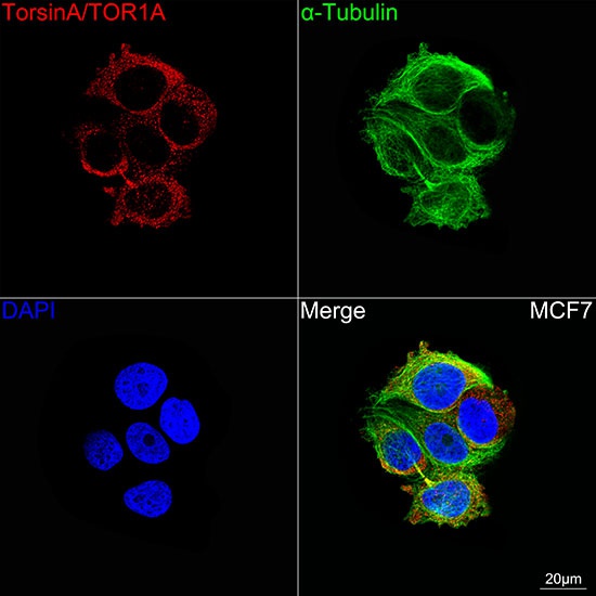
Confocal imaging of MCF7 cells using TorsinA/TOR1A Rabbit mAb (CAB9579, dilution 1:200) followed by a further incubation with Cy3 Goat Anti-Rabbit IgG (H+L) (CABS007, dilution 1:500) (Red). The cells were counterstained with α-Tubulin Mouse mAb (AC012, dilution 1:400) followed by incubation with ABflo® 488-conjugated Goat Anti-Mouse IgG (H+L) Ab (CABS076, dilution 1:500) (Green). DAPI was used for nuclear staining (Blue). Objective: 100x.