The WAPL Monoclonal Antibody (CAB5923) is a high-quality antibody developed for reliable detection and analysis of target proteins. This antibody, produced in rabbits, is specifically reactive with human WAPL samples and has been validated for use in applications such as Western blotting.The WAPL protein is crucial for ensuring the accurate distribution of genetic material during cell division, making it a key player in maintaining genomic stability. Dysregulation of WAPL function has been implicated in diseases such as cancer, where abnormal cell division can lead to uncontrolled growth and tumor formation.
This antibody is validated for use in WB, IF/ICC, IP, ELISA applications and has demonstrated reactivity against Human, Mouse samples.
Product Name:
WAPL Monoclonal Antibody
SKU:
CAB5923
Size:
20μL, 100μL
Reactivity:
Human, Mouse
Clone Number:
ARC2088
Conjugate:
Unconjugated
Immunogen:
Synthetic peptide. This information is considered to be commercially sensitive.
Involved in several processes, including negative regulation of DNA replication; negative regulation of chromatin binding activity; and regulation of sister chromatid cohesion. Located in several cellular components, including Golgi apparatus; intercellular bridge; and microtubule cytoskeleton.
Purification Method
Affinity purification
Gene ID
23063
Buffer Information
Store at -20℃. Avoid freeze / thaw cycles. Buffer: PBS containing 50% glycerol and 0.05% BSA, preserved with proclin300 or sodium azide, pH 7.3.
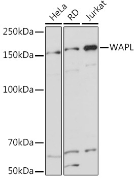
Western blot analysis of various lysates using WAPL Rabbit mAb (CAB5923) at 1:1000 dilution. Secondary antibody: HRP-conjugated Goat anti-Rabbit IgG (H+L) (CABS014) at 1:10000 dilution. Lysates/proteins: 25μg per lane. Blocking buffer: 3% nonfat dry milk in TBST. Detection: ECL Basic Kit (AbGn00020). Exposure time: 1s.
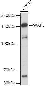
Western blot analysis of lysates from C2C12 cells, using WAPL Rabbit mAb (CAB5923) at 1:1000 dilution. Secondary antibody: HRP-conjugated Goat anti-Rabbit IgG (H+L) (CABS014) at 1:10000 dilution. Lysates/proteins: 25μg per lane. Blocking buffer: 3% nonfat dry milk in TBST. Detection: ECL Basic Kit (AbGn00020). Exposure time: 10s.
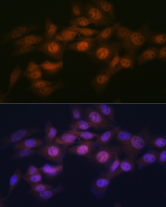
Immunofluorescence analysis of NIH-3T3 cells using WAPL Rabbit mAb (CAB5923) at dilution of 1:100 (40x lens). Secondary antibody: Cy3-conjugated Goat anti-Rabbit IgG (H+L) (CABS007) at 1:500 dilution. Blue: DAPI for nuclear staining.
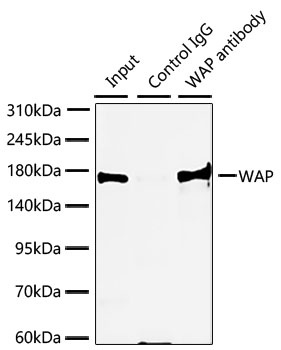
Immunoprecipitation of WAPL from 300 µg extracts of HeLa cells was performed using 3 µg of WAPL Rabbit mAb (CAB5923). Rabbit Control IgG (AC005) was used to precipitate the Control IgG sample. IP samples were eluted with 1X Laemmli Buffer. The Input lane represents 10% of the total input. Western blot analysis of immunoprecipitates was conducted using WAPL Rabbit mAb (CAB5923) at a dilution of 1:1000.