The AP3D1 Antibody (CAB13058) is a high-quality antibody developed for reliable detection and analysis of target proteins. This antibody, produced in rabbits, exhibits high reactivity with human samples and has been validated for use in Western blot applications. By specifically binding to AP3D1, researchers can accurately detect and analyze this important protein in a variety of cell types, making it an essential tool for studies in cell biology and molecular biology.AP3D1 is a crucial component of the AP-3 protein complex, which plays a critical role in sorting cargo proteins for delivery to lysosomes and related organelles.
This antibody is validated for use in WB, ELISA applications and has demonstrated reactivity against Human, Mouse samples.
Product Name:
AP3D1 Antibody
SKU:
CAB13058
Size:
20μL, 100μL
Reactivity:
Human, Mouse
Conjugate:
Unconjugated
Immunogen:
Recombinant protein (or fragment).This information is considered to be commercially sensitive.
The protein encoded by this gene is a subunit of the AP3 adaptor-like complex, which is not clathrin-associated, but is associated with the golgi region, as well as more peripheral structures. The AP-3 complex facilitates the budding of vesicles from the golgi membrane, and may be directly involved in trafficking to lysosomes. This subunit is implicated in intracellular biogenesis and trafficking of pigment granules, and possibly platelet dense granules and neurotransmitter vesicles. Defects in this gene are a cause of a new type of Hermansky-Pudlak syndrome.
Purification Method
Affinity purification
Gene ID
8943
RRID
AB_2759906
Buffer Information
Store at -20℃. Avoid freeze / thaw cycles. Buffer: PBS with 0.01% thimerosal,50% glycerol,pH7.3.
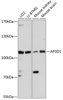
Western blot analysis of various lysates using AP3D1 Rabbit pAb (CAB13058) at 1:3000 dilution. Secondary antibody: HRP-conjugated Goat anti-Rabbit IgG (H+L) (CABS014) at 1:10000 dilution. Lysates/proteins: 25μg per lane. Blocking buffer: 3% nonfat dry milk in TBST. Detection: ECL Basic Kit (AbGn00020). Exposure time: 90s.