The APEH Antibody (CAB5893) is a high-quality antibody developed for reliable detection and analysis of target proteins. The antibody is produced in rabbits and exhibits high reactivity towards human samples, making it suitable for use in Western blot applications. By targeting the APEH protein, this antibody allows for the precise detection and analysis of APEH in various cell types, making it an essential component for studies focused on enzymatic activity and its role in cellular processes.APEH, also known as acylpeptide hydrolase, plays a critical role in the regulation of protein degradation and turnover within the cell.
This antibody is validated for use in WB, IP, ELISA applications and has demonstrated reactivity against Human, Mouse, Rat samples.
Product Name:
APEH Antibody
SKU:
CAB5893
Size:
20μL, 100μL
Reactivity:
Human, Mouse, Rat
Conjugate:
Unconjugated
Immunogen:
Recombinant protein (or fragment).This information is considered to be commercially sensitive.
This gene encodes the enzyme acylpeptide hydrolase, which catalyzes the hydrolysis of the terminal acetylated amino acid preferentially from small acetylated peptides. The acetyl amino acid formed by this hydrolase is further processed to acetate and a free amino acid by an aminoacylase. This gene is located within the same region of chromosome 3 (3p21) as the aminoacylase gene, and deletions at this locus are also associated with a decrease in aminoacylase activity. The acylpeptide hydrolase is a homotetrameric protein of 300 kDa with each subunit consisting of 732 amino acid residues. It can play an important role in destroying oxidatively damaged proteins in living cells. Deletions of this gene locus are found in various types of carcinomas, including small cell lung carcinoma and renal cell carcinoma.
Purification Method
Affinity purification
Gene ID
327
RRID
AB_2766641
Buffer Information
Store at -20℃. Avoid freeze / thaw cycles. Buffer: PBS containing 50% glycerol, preserved with proclin300 or sodium azide, pH 7.3.
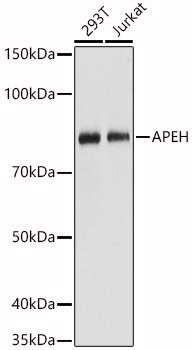
Western blot analysis of various lysates, using [KO Validated] APEH Rabbit pAb (CAB5893) at 1:1000 dilution. Secondary antibody: HRP-conjugated Goat anti-Rabbit IgG (H+L) (CABS014) at 1:10000 dilution. Lysates/proteins: 25μg per lane. Blocking buffer: 3% nonfat dry milk in TBST. Detection: ECL Basic Kit (AbGn00020). Exposure time: 1s.
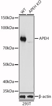
Western blot analysis of lysates from wild type(WT) and APEH knockout (KO) 293T(KO) cells, using [KO Validated] APEH Rabbit pAb (CAB5893) at 1:1000 dilution. Secondary antibody: HRP-conjugated Goat anti-Rabbit IgG (H+L) (CABS014) at 1:10000 dilution. Lysates/proteins: 25μg per lane. Blocking buffer: 3% nonfat dry milk in TBST. Detection: ECL Basic Kit (AbGn00020). Exposure time: 1s.