The APIP Antibody (CAB7102) is a high-quality antibody developed for reliable detection and analysis of target proteins. APIP is known for its role in apoptosis and its potential as a therapeutic target for cancer treatment. This antibody, produced in rabbits, is highly specific and reactive with human samples, making it ideal for Western blot applications. By targeting the APIP protein, researchers can effectively analyze and detect its expression in various cell types, providing valuable insights into apoptosis pathways and potential treatment strategies for cancer and other diseases.
This antibody is validated for use in WB, IHC-P, ELISA applications and has demonstrated reactivity against Human, Mouse, Rat samples.
Product Name:
APIP Antibody
SKU:
CAB7102
Size:
20μL, 100μL
Reactivity:
Human, Mouse, Rat
Conjugate:
Unconjugated
Immunogen:
Recombinant protein (or fragment).This information is considered to be commercially sensitive.
Enables identical protein binding activity; methylthioribulose 1-phosphate dehydratase activity; and zinc ion binding activity. Involved in several processes, including L-methionine salvage from methylthioadenosine; protein homotetramerization; and pyroptosis. Located in cytoplasm.
Purification Method
Affinity purification
Gene ID
51074
RRID
AB_2767657
Buffer Information
Store at -20℃. Avoid freeze / thaw cycles. Buffer: PBS containing 50% glycerol, preserved with proclin300 or sodium azide, pH 7.3.
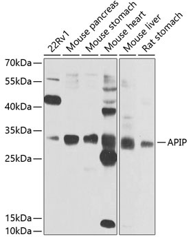
Western blot analysis of various lysates using APIP Rabbit pAb (CAB7102) at 1:1000 dilution. Secondary antibody: HRP-conjugated Goat anti-Rabbit IgG (H+L) (CABS014) at 1:10000 dilution. Lysates/proteins: 25μg per lane. Blocking buffer: 3% nonfat dry milk in TBST. Detection: ECL Enhanced Kit (AbGn00021). Exposure time: 10s.
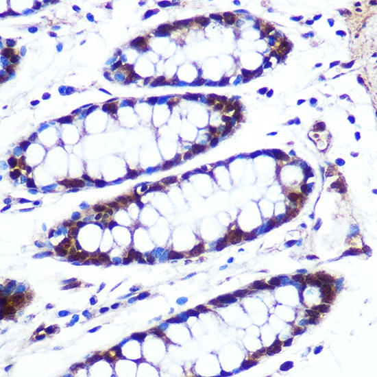
Immunohistochemistry analysis of paraffin-embedded Human colon using APIP Rabbit pAb (CAB7102) at dilution of 1:100 (40x lens). Microwave antigen retrieval performed with 0.01M PBS Buffer (pH 7.2) prior to IHC staining.