The APLP1 Antibody (CAB6870) is a high-quality antibody developed for reliable detection and analysis of target proteins. This antibody, produced in rabbits, exhibits high reactivity with human samples and has been validated for use in Western blot applications.APLP1 is a member of the amyloid precursor protein (APP) family and is known to interact with proteins involved in Alzheimer's disease pathology. Research suggests that APLP1 may play a role in synaptic plasticity and neuronal survival, making it a promising target for investigations into neurodegenerative disorders.
This antibody is validated for use in WB, IHC-P, IF/ICC, ELISA applications and has demonstrated reactivity against Human, Mouse, Rat samples.
Product Name:
APLP1 Antibody
SKU:
CAB6870
Size:
20μL, 100μL
Reactivity:
Human, Mouse, Rat
Conjugate:
Unconjugated
Immunogen:
Recombinant protein (or fragment).This information is considered to be commercially sensitive.
Recommended starting concentration is 1 μg/mL. Please optimize the concentration based on your specific assay requirements.
Synonyms:
APLP, APLP1
Positive Sample:
Mouse brain, Rat brain
Cellular Localization:
Cell Membrane, Cytoplasm, Single-Pass Type I Membrane Protein.
Calculated MW:
72kDa
Observed MW:
72kDa
This gene encodes a member of the highly conserved amyloid precursor protein gene family. The encoded protein is a membrane-associated glycoprotein that is cleaved by secretases in a manner similar to amyloid beta A4 precursor protein cleavage. This cleavage liberates an intracellular cytoplasmic fragment that may act as a transcriptional activator. The encoded protein may also play a role in synaptic maturation during cortical development. Alternatively spliced transcript variants encoding different isoforms have been described.
Purification Method
Affinity purification
Gene ID
333
RRID
AB_2767430
Buffer Information
Store at -20℃. Avoid freeze / thaw cycles. Buffer: PBS containing 50% glycerol, preserved with proclin300 or sodium azide, pH 7.3.
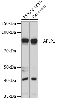
Western blot analysis of various lysates using APLP1 Rabbit pAb (CAB6870) at 1:1000 dilution. Secondary antibody: HRP-conjugated Goat anti-Rabbit IgG (H+L) (CABS014) at 1:10000 dilution. Lysates/proteins: 25μg per lane. Blocking buffer: 3% nonfat dry milk in TBST. Detection: ECL Basic Kit (AbGn00020). Exposure time: 10s.
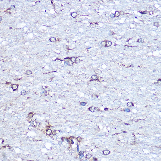
Immunohistochemistry analysis of paraffin-embedded Rat brain using APLP1 Rabbit pAb (CAB6870) at dilution of 1:100 (40x lens). Microwave antigen retrieval performed with 0.01M Tris/EDTA Buffer (pH 9.0) prior to IHC staining.
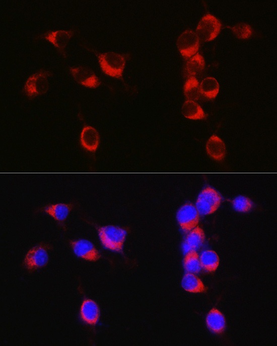
Immunofluorescence analysis of Neuro-2a cells using APLP1 Rabbit pAb (CAB6870) at dilution of 1:50 (40x lens). Secondary antibody: Cy3-conjugated Goat anti-Rabbit IgG (H+L) (CABS007) at 1:500 dilution. Blue: DAPI for nuclear staining.
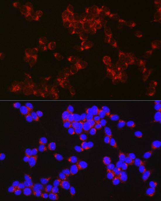
Immunofluorescence analysis of SH-SY5Y cells using APLP1 Rabbit pAb (CAB6870) at dilution of 1:50 (40x lens). Secondary antibody: Cy3-conjugated Goat anti-Rabbit IgG (H+L) (CABS007) at 1:500 dilution. Blue: DAPI for nuclear staining.