The APP Antibody (CAB11019) is a high-quality antibody developed for reliable detection and analysis of target proteins. APP is a transmembrane protein that plays a key role in the generation of amyloid beta peptides, which are implicated in the pathogenesis of Alzheimer's disease.This antibody, generated in rabbits, has been validated for use in various applications, including Western blotting and immunohistochemistry. It selectively binds to the APP protein, enabling researchers to accurately detect and analyze its expression in samples from various species, including human, mouse, and rat.
This antibody is validated for use in WB, IF/ICC, ELISA applications and has demonstrated reactivity against Human, Mouse, Rat samples.
Product Name:
APP Antibody
SKU:
CAB11019
Size:
20μL, 100μL
Reactivity:
Human, Mouse, Rat
Conjugate:
Unconjugated
Immunogen:
Synthetic peptide. This information is considered to be commercially sensitive.
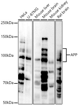
HeLa, U-87MG, Mouse liver, Mouse brain, Mouse kidney, Rat brain
Cellular Localization:
Membrane, Single-Pass Type I Membrane Protein, Clathrin-Coated Pit.
Calculated MW:
87kDa
Observed MW:
100-140kDa
This gene encodes a cell surface receptor and transmembrane precursor protein that is cleaved by secretases to form a number of peptides. Some of these peptides are secreted and can bind to the acetyltransferase complex APBB1/TIP60 to promote transcriptional activation, while others form the protein basis of the amyloid plaques found in the brains of patients with Alzheimer disease. In addition, two of the peptides are antimicrobial peptides, having been shown to have bacteriocidal and antifungal activities. Mutations in this gene have been implicated in autosomal dominant Alzheimer disease and cerebroarterial amyloidosis (cerebral amyloid angiopathy). Multiple transcript variants encoding several different isoforms have been found for this gene.
Purification Method
Affinity purification
Gene ID
351
RRID
AB_2758368
Buffer Information
Store at -20℃. Avoid freeze / thaw cycles. Buffer: PBS containing 50% glycerol, preserved with proclin300 or sodium azide, pH 7.3.
Western blot analysis of various lysates, using APP Rabbit pAb (CAB11019) at 1:1000 dilution. Secondary antibody: HRP-conjugated Goat anti-Rabbit IgG (H+L) (CABS014) at 1:10000 dilution. Lysates/proteins: 25μg per lane. Blocking buffer: 3% nonfat dry milk in TBST. Detection: ECL Basic Kit (AbGn00020). Exposure time: 30s.
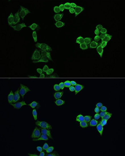
Immunofluorescence analysis of HeLa cells using APP Rabbit pAb (CAB11019) at dilution of 1:100. Blue: DAPI for nuclear staining.
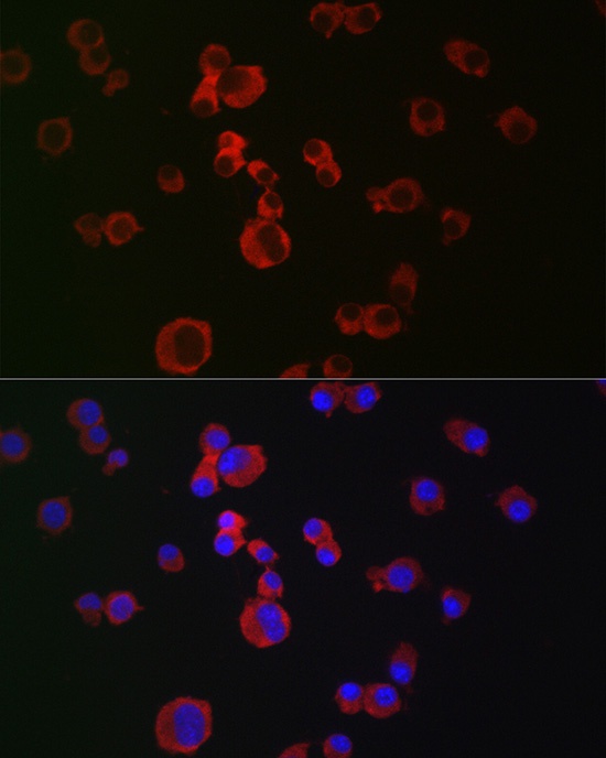
Immunofluorescence analysis of Neuro-2a cells using APP Rabbit pAb (CAB11019) at dilution of 1:100 (40x lens). Secondary antibody: Cy3-conjugated Goat anti-Rabbit IgG (H+L) (CABS007) at 1:500 dilution. Blue: DAPI for nuclear staining.
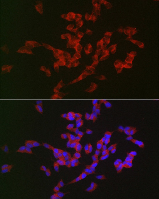
Immunofluorescence analysis of SH-SY5Y cells using APP Rabbit pAb (CAB11019) at dilution of 1:100 (40x lens). Secondary antibody: Cy3-conjugated Goat anti-Rabbit IgG (H+L) (CABS007) at 1:500 dilution. Blue: DAPI for nuclear staining.