The Aquaporin-1 (AQP1) Monoclonal Antibody (CAB4195) is a high-quality antibody developed for reliable detection and analysis of target proteins. This gene encodes a small integral membrane protein with six bilayer spanning domains that functions as a water channel protein. This protein permits passive transport of water along an osmotic gradient. This gene is a possible candidate for disorders involving imbalance in ocular fluid movement. RRID AB_2863207 Gene ID 358 Swiss Prot Synonym CO; CHIP28; AQP-CHIP; Aquaporin-1 (AQP1)
This antibody is validated for use in WB, IHC-P, ELISA, IF-P applications and has demonstrated reactivity against Human, Mouse, Rat samples.
Product Name:
Aquaporin-1 (AQP1) Monoclonal Antibody
SKU:
CAB4195
Size:
100μL, 20μL
Reactivity:
Human, Mouse, Rat
Clone Number:
ARC0925
Conjugate:
Unconjugated
Immunogen:
Synthetic peptide. This information is considered to be commercially sensitive.
Tested Applications:
WBIHC-PELISAIF-P
Recommended Dilution:
WB
1:1000 - 1:6000
IF-P
1:100 - 1:1000
IHC-P
1:500 - 1:2000
ELISA
Recommended starting concentration is 1 μg/mL. Please optimize the concentration based on your specific assay requirements.
Synonyms:
CO, CHIP28, AQP-CHIP, Aquaporin-1 (AQP1)
Positive Sample:
Mouse liver, Rat spleen, Rat kidney
Cellular Localization:
Cell Membrane, Multi-Pass Membrane Protein.
Calculated MW:
29kDa
Observed MW:
29kDa
This gene encodes a small integral membrane protein with six bilayer spanning domains that functions as a water channel protein. This protein permits passive transport of water along an osmotic gradient. This gene is a possible candidate for disorders involving imbalance in ocular fluid movement. RRID AB_2863207 Gene ID 358 Swiss Prot Synonym CO; CHIP28; AQP-CHIP; Aquaporin-1 (AQP1)
Purification Method:
Affinity purification
Gene ID:
358
RRID:
AB_2863207
Buffer Information:
Store at -20℃. Avoid freeze / thaw cycles. Buffer: PBS containing 50% glycerol and 0.05% BSA, preserved with proclin300 or sodium azide, pH 7.3.
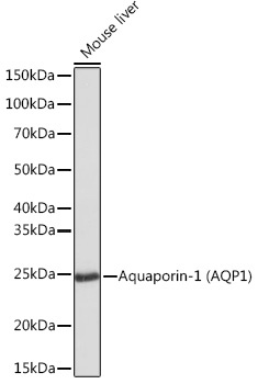
Western blot analysis of lysates from Mouse liver, using Aquaporin-1 (Aquaporin-1 (AQP1)) Rabbit mAb (CAB4195) at 1:1000 dilution. Secondary antibody: HRP-conjugated Goat anti-Rabbit IgG (H+L) (AS014) at 1:10000 dilution. Lysates/proteins: 25μg per lane. Blocking buffer: 3% nonfat dry milk in TBST. Detection: ECL Basic Kit (AbGn00020). Exposure time: 1s.
Western blot analysis of various lysates using Aquaporin-1 (Aquaporin-1 (AQP1)) Rabbit mAb (CAB4195) at 1:1000 dilution. Secondary antibody: HRP-conjugated Goat anti-Rabbit IgG (H+L) (AS014) at 1:10000 dilution. Lysates/proteins: 25μg per lane. Blocking buffer: 3% nonfat dry milk in TBST. Detection: ECL Basic Kit (AbGn00020). Exposure time: 10s.
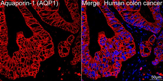
Confocal imaging of paraffin-embedded Human colon cancer tissue using Aquaporin-1 (AQP1) Rabbit mAb (CAB4195, dilution 1:100) followed by a further incubation with Cy3 Goat Anti-Rabbit IgG (H+L) (AS007, dilution 1:500) (Red). DAPI was used for nuclear staining (Blue). High pressure antigen retrieval performed with 0.01M Citrate Buffer (pH 6.0) prior to IF staining. Objective: 40x.
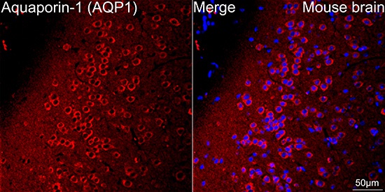
Confocal imaging of paraffin-embedded Mouse brain tissue using Aquaporin-1 (AQP1) Rabbit mAb (CAB4195, dilution 1:100) followed by a further incubation with Cy3 Goat Anti-Rabbit IgG (H+L) (AS007, dilution 1:500) (Red). DAPI was used for nuclear staining (Blue). Microwave antigen retrieval performed with 0.01M Citrate Buffer (pH 6.0) prior to IF staining. Objective: 40x.


 ELISA Kit (MOEB0465)")
")
