The ARAF Antibody (CAB16346) is a high-quality antibody developed for reliable detection and analysis of target proteins. This antibody, generated in rabbits, exhibits high reactivity with human samples and is validated for use in Western blot applications. By specifically binding to the ARAF protein, researchers can easily detect and analyze its expression in a variety of cell types, making it an excellent choice for studies in cancer biology and signal transduction research.ARAF plays a crucial role in cell growth, proliferation, and differentiation, making it a key player in various cellular processes.
This antibody is validated for use in WB, IF/ICC, ELISA applications and has demonstrated reactivity against Human, Mouse, Rat samples.
Product Name:
ARAF Antibody
SKU:
CAB16346
Size:
20μL, 100μL
Reactivity:
Human, Mouse, Rat
Conjugate:
Unconjugated
Immunogen:
Recombinant protein (or fragment).This information is considered to be commercially sensitive.
Recommended starting concentration is 1 μg/mL. Please optimize the concentration based on your specific assay requirements.
Synonyms:
PKS2, A-RAF, ARAF1, RAFA1, ARAF
Cellular Localization:
Cytosol, Mitochondrion.
Calculated MW:
68kDa
Observed MW:
Refertofigures
Enables protein serine/threonine kinase activity. Involved in negative regulation of apoptotic process; regulation of TOR signaling; and regulation of cellular protein metabolic process. Predicted to be active in cytosol and mitochondrion. Biomarker of high grade glioma.
Purification Method
Affinity purification
Gene ID
369
RRID
AB_2768411
Buffer Information
Store at -20℃. Avoid freeze / thaw cycles. Buffer: PBS containing 50% glycerol, preserved with proclin300 or sodium azide, pH 7.3.
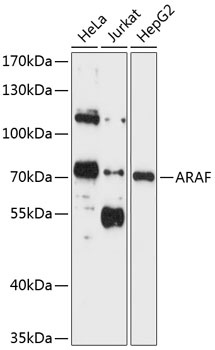
Western blot analysis of various lysates using ARAF Rabbit pAb (CAB16346) at 1:1000 dilution._Secondary antibody: HRP-conjugated Goat anti-Rabbit IgG (H+L) (CABS014) at 1:10000 dilution._Lysates/proteins: 25μg per lane._Blocking buffer: 3% nonfat dry milk in TBST._Detection: ECL Enhanced Kit (AbGn00021)._Exposure time: 5s.
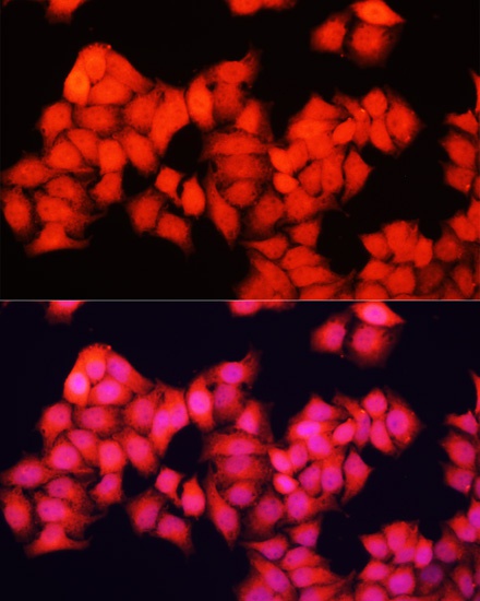
Immunofluorescence analysis of HeLa cells using ARAF Rabbit pAb (CAB16346) at dilution of 1:100. Secondary antibody: Cy3-conjugated Goat anti-Rabbit IgG (H+L) (CABS007) at 1:500 dilution. Blue: DAPI for nuclear staining.