The AREG Antibody (CAB12680) is a high-quality antibody developed for reliable detection and analysis of target proteins. This antibody, produced in rabbits, exhibits high specificity and sensitivity to AREG in human samples, making it ideal for use in immunoassays such as Western blotting.AREG is known to regulate various cellular processes, including cell growth, migration, and survival, making it a promising target for cancer research and therapy development.
This antibody is validated for use in IF/ICC, ELISA applications and has demonstrated reactivity against Mouse samples.
Product Name:
AREG Antibody
SKU:
CAB12680
Size:
20μL, 100μL
Reactivity:
Mouse
Conjugate:
Unconjugated
Immunogen:
Recombinant protein (or fragment).This information is considered to be commercially sensitive.
Recommended starting concentration is 1 μg/mL. Please optimize the concentration based on your specific assay requirements.
Synonyms:
AR, SDGF, AREGB, CRDGF, AREG
Cellular Localization:
Membrane, Single-Pass Membrane Protein.
Calculated MW:
28kDa
Observed MW:
Refertofigures
The protein encoded by this gene is a member of the epidermal growth factor family. It is an autocrine growth factor as well as a mitogen for astrocytes, Schwann cells and fibroblasts. It is related to epidermal growth factor (EGF) and transforming growth factor alpha (TGF-alpha). The protein interacts with the EGF/TGF-alpha receptor to promote the growth of normal epithelial cells, and it inhibits the growth of certain aggressive carcinoma cell lines. It also functions in mammary gland, oocyte and bone tissue development. This gene is associated with a psoriasis-like skin phenotype, and is also associated with other pathological disorders, including various types of cancers and inflammatory conditions.
Purification Method
Affinity purification
Gene ID
374
RRID
AB_2759526
Buffer Information
Store at -20℃. Avoid freeze / thaw cycles. Buffer: Buffer: PBS containing 50% glycerol, preserved with proclin300 or sodium azide, pH 7.3.
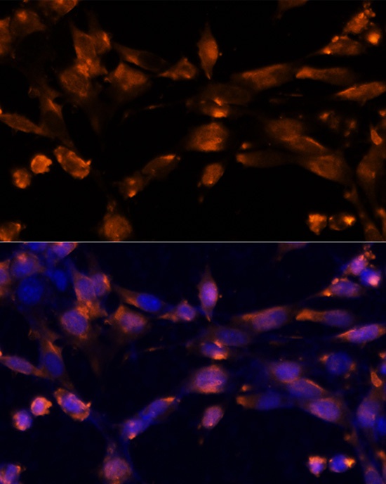
Immunofluorescence analysis of NIH-3T3 cells using AREG Rabbit pAb (CAB12680) at dilution of 1:100 (40x lens). Secondary antibody: Cy3-conjugated Goat anti-Rabbit IgG (H+L) (CABS007) at 1:500 dilution. Blue: DAPI for nuclear staining.