The ASCC3 Antibody (CAB7960) is a high-quality antibody developed for reliable detection and analysis of target proteins. This antibody, produced in rabbits, is highly specific to ASCC3 in human samples and is validated for use in Western blot applications. By binding to ASCC3, this antibody allows for the detection and analysis of the protein in various cell types, making it an ideal choice for studies in molecular biology and cancer research.ASCC3, also known as Activating Signal Cointegrator 1 Complex Subunit 3, plays a critical role in maintaining genomic integrity by participating in DNA repair mechanisms.
This antibody is validated for use in WB, IF/ICC, ELISA applications and has demonstrated reactivity against Human, Mouse samples.
Product Name:
ASCC3 Antibody
SKU:
CAB7960
Size:
20μL, 100μL
Reactivity:
Human, Mouse
Conjugate:
Unconjugated
Immunogen:
Recombinant protein (or fragment).This information is considered to be commercially sensitive.
Recommended starting concentration is 1 μg/mL. Please optimize the concentration based on your specific assay requirements.
Synonyms:
RNAH, HELIC1, ASC1p200, ASCC3
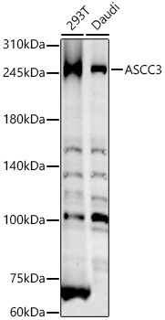
Positive Sample:
293T, Daudi
Cellular Localization:
Cytoplasm, Nucleus.
Calculated MW:
251kDa
Observed MW:
251kDa
This gene encodes a protein that belongs to a family of helicases that are involved in the ATP-dependent unwinding of nucleic acid duplexes. The encoded protein is the largest subunit of the activating signal cointegrator 1 complex that is involved in DNA repair and resistance to alkylation damage. Alternate splicing results in multiple transcript variants.
Purification Method
Affinity purification
Gene ID
10973
RRID
AB_2768471
Buffer Information
Store at -20℃. Avoid freeze / thaw cycles. Buffer: PBS containing 50% glycerol, preserved with proclin300 or sodium azide, pH 7.3.
Western blot analysis of various lysates, using ASCC3 Rabbit pAb (CAB7960) at 1:800 dilution. Secondary antibody: HRP-conjugated Goat anti-Rabbit IgG (H+L) (CABS014) at 1:10000 dilution. Lysates/proteins: 25μg per lane. Blocking buffer: 3% nonfat dry milk in TBST. Detection: ECL Basic Kit (AbGn00020). Exposure time: 50s.
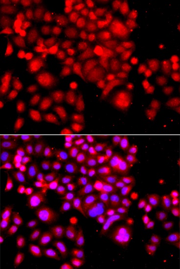
Immunofluorescence analysis of A549 cells using ASCC3 Rabbit pAb (CAB7960). Secondary antibody: Cy3-conjugated Goat anti-Rabbit IgG (H+L) (CABS007) at 1:500 dilution. Blue: DAPI for nuclear staining.