The ASH2L Monoclonal Antibody (CAB4892) is a high-quality antibody developed for reliable detection and analysis of target proteins. This high-quality antibody, generated using rabbit monoclonal technology, exhibits exceptional specificity and sensitivity in detecting ASH2L in various experimental settings.ASH2L is known to be a subunit of the SET1/MLL histone methyltransferase complexes, playing a role in epigenetic regulation and transcriptional activation. Dysregulation of ASH2L has been linked to various diseases, including cancer and developmental disorders, making it a promising target for therapeutic interventions.
This antibody is validated for use in WB, IHC-P, IF/ICC, ELISA, IF-P applications and has demonstrated reactivity against Human, Mouse, Rat samples.
Product Name:
ASH2L Monoclonal Antibody
SKU:
CAB4892
Size:
20μL, 100μL
Reactivity:
Human, Mouse, Rat
Clone Number:
ARC0326
Conjugate:
Unconjugated
Immunogen:
Synthetic peptide. This information is considered to be commercially sensitive.
Recommended starting concentration is 1 μg/mL. Please optimize the concentration based on your specific assay requirements.
Synonyms:
ASH2, Bre2, ASH2L1, ASH2L2, ASH2L
Positive Sample:
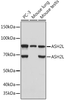
PC-3, Mouse lung, Mouse testis
Cellular Localization:
Nucleus.
Calculated MW:
69kDa
Observed MW:
69kDa/85kDa
Enables beta-catenin binding activity and transcription cis-regulatory region binding activity. Contributes to histone methyltransferase activity (H3-K4 specific). Involved in histone H3-K4 methylation; positive regulation of cell population proliferation; and response to estrogen. Acts upstream of or within cellular response to DNA damage stimulus. Located in nucleus. Part of MLL3/4 complex and Set1C/COMPASS complex.
Purification Method
Affinity purification
Gene ID
9070
RRID
AB_2863377
Buffer Information
Store at -20℃. Avoid freeze / thaw cycles. Buffer: PBS containing 50% glycerol and 0.05% BSA, preserved with proclin300 or sodium azide, pH 7.3.
Western blot analysis of various lysates using ASH2L Rabbit mAb (CAB4892) at 1:1000 dilution. Secondary antibody: HRP-conjugated Goat anti-Rabbit IgG (H+L) (CABS014) at 1:10000 dilution. Lysates/proteins: 25μg per lane. Blocking buffer: 3% nonfat dry milk in TBST. Detection: ECL Basic Kit (AbGn00020). Exposure time: 5s.
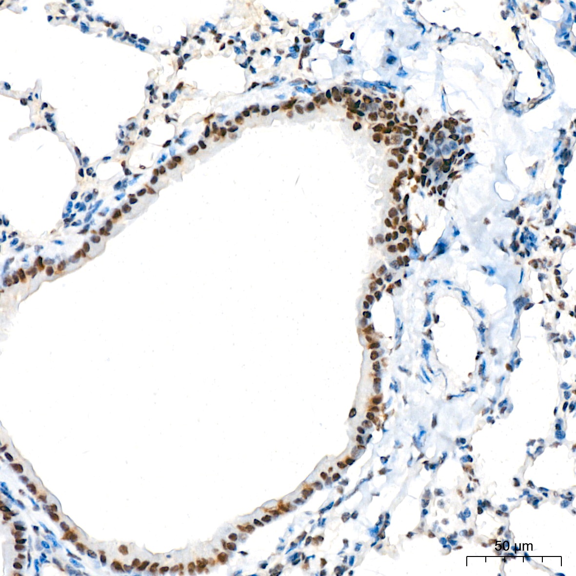
Immunohistochemistry analysis of paraffin-embedded Mouse lung tissue using ASH2L Rabbit mAb (CAB4892) at a dilution of 1:200 (40x lens). High pressure antigen retrieval was performed with 0.01 M citrate buffer (pH 6.0) prior to IHC staining.
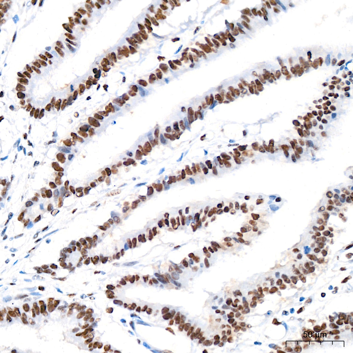
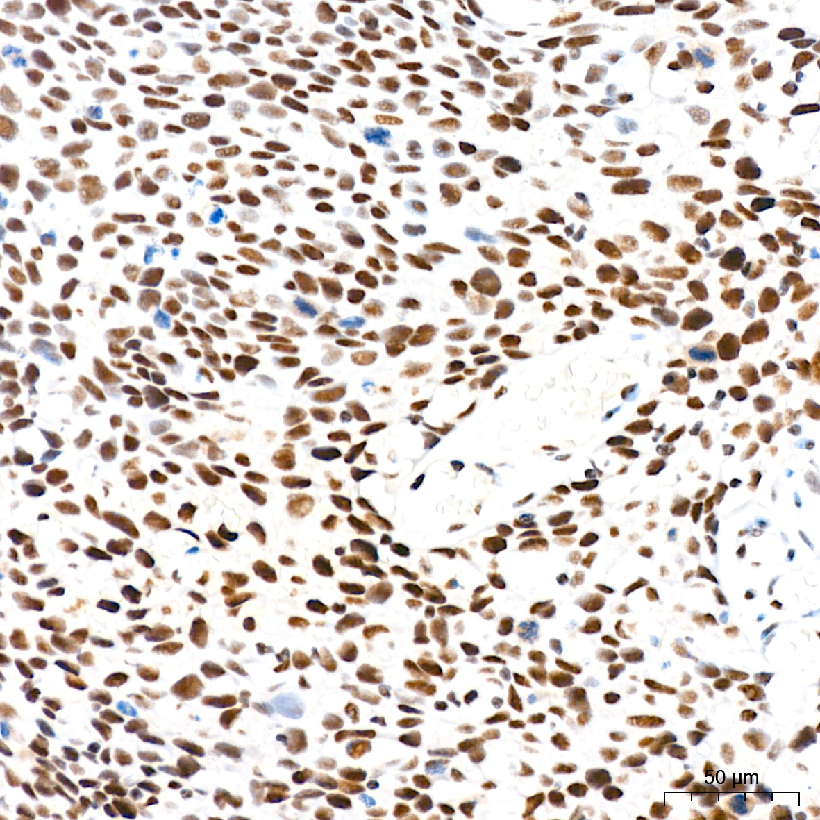
Immunohistochemistry analysis of paraffin-embedded Human colon carcinoma tissue using ASH2L Rabbit mAb (CAB4892) at a dilution of 1:200 (40x lens). High pressure antigen retrieval was performed with 0.01 M citrate buffer (pH 6.0) prior to IHC staining.
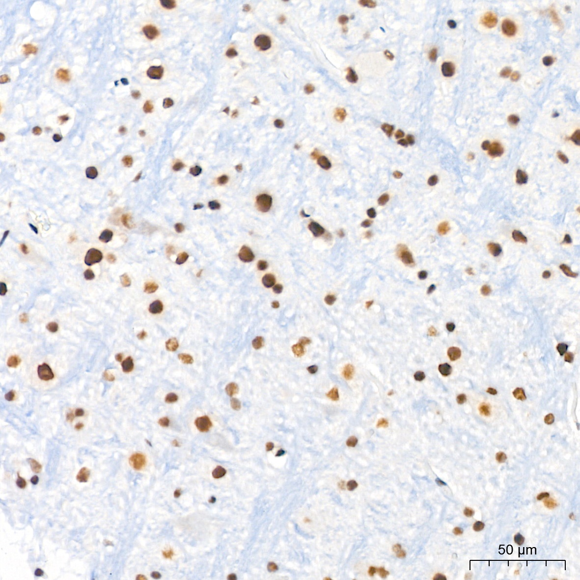
Immunohistochemistry analysis of paraffin-embedded Mouse brain tissue using ASH2L Rabbit mAb (CAB4892) at a dilution of 1:200 (40x lens). High pressure antigen retrieval was performed with 0.01 M citrate buffer (pH 6.0) prior to IHC staining.
Immunohistochemistry analysis of paraffin-embedded Human cervix cancer tissue using ASH2L Rabbit mAb (CAB4892) at a dilution of 1:200 (40x lens). High pressure antigen retrieval was performed with 0.01 M citrate buffer (pH 6.0) prior to IHC staining.
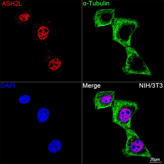
Confocal imaging of NIH/3T3 cells using ASH2L Rabbit mAb (CAB4892, dilution 1:100) followed by a further incubation with Cy3 Goat Anti-Rabbit IgG (H+L) (CABS007, dilution 1:500) (Red). The cells were counterstained with α-Tubulin Mouse mAb (AC012, dilution 1:400) followed by incubation with ABflo® 488-conjugated Goat Anti-Mouse IgG (H+L) Ab (CABS076, dilution 1:500) (Green). DAPI was used for nuclear staining (Blue). Objective: 100x.
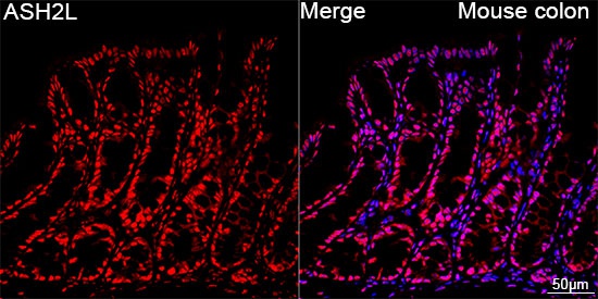
Confocal imaging of paraffin-embedded Mouse colon tissue using ASH2L Rabbit mAb (CAB4892, dilution 1:100) followed by a further incubation with Cy3 Goat Anti-Rabbit IgG (H+L) (CABS007, dilution 1:500) (Red). DAPI was used for nuclear staining (Blue). Objective: 40x. Perform high pressure antigen retrieval with 0.01 M citrate buffer (pH 6.0) prior to IF staining.