The ATXN10 Antibody (CAB4586) is a high-quality antibody developed for reliable detection and analysis of target proteins. This antibody, produced in rabbits, is highly specific to human samples and has been validated for use in Western blot applications. By targeting the Ataxin-10 protein, this antibody enables researchers to detect and analyze its expression in a variety of cell types, making it an essential tool for studies in neurology and genetic diseases.Ataxin-10 is a key player in the pathogenesis of SCA10, making it a promising target for therapeutic interventions aimed at treating this debilitating condition.
This antibody is validated for use in WB, IF/ICC, ELISA applications and has demonstrated reactivity against Human, Mouse, Rat samples.
Product Name:
ATXN10 Antibody
SKU:
CAB4586
Size:
20μL, 100μL
Reactivity:
Human, Mouse, Rat
Conjugate:
Unconjugated
Immunogen:
Recombinant protein (or fragment).This information is considered to be commercially sensitive.
This gene encodes a protein that may function in neuron survival, neuron differentiation, and neuritogenesis. These roles may be carried out via activation of the mitogen-activated protein kinase cascade. Expansion of an ATTCT repeat from 9-32 copies to 800-4500 copies in an intronic region of this locus has been associated with spinocerebellar ataxia, type 10. Alternatively spliced transcript variants have been described.
Purification Method
Affinity purification
Gene ID
25814
RRID
AB_2765770
Buffer Information
Store at -20℃. Avoid freeze / thaw cycles. Buffer: PBS containing 50% glycerol, preserved with proclin300 or sodium azide, pH 7.3.
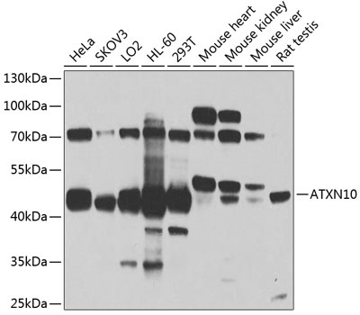
Western blot analysis of various lysates using ATXN10 Rabbit pAb (CAB4586) at 1:1000 dilution. Secondary antibody: HRP-conjugated Goat anti-Rabbit IgG (H+L) (CABS014) at 1:10000 dilution. Lysates/proteins: 25μg per lane. Blocking buffer: 3% nonfat dry milk in TBST. Detection: ECL Basic Kit (AbGn00020). Exposure time: 90s.
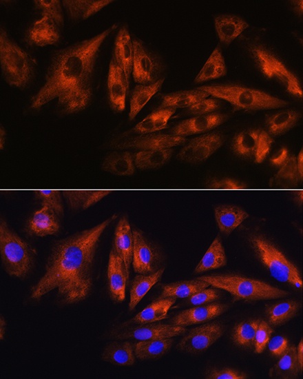
Immunofluorescence analysis of H9C2 cells using ATXN10 Rabbit pAb (CAB4586) at dilution of 1:100. Secondary antibody: Cy3-conjugated Goat anti-Rabbit IgG (H+L) (CABS007) at 1:500 dilution. Blue: DAPI for nuclear staining.