The ATF3 Antibody (CAB13469) is a high-quality antibody developed for reliable detection and analysis of target proteins. This polyclonal antibody, generated in rabbits, exhibits high reactivity with human samples and is validated for use in Western blot applications. By specifically binding to the ATF3 protein, this antibody enables precise detection and analysis in a variety of cell types, making it well-suited for investigations in molecular biology and disease research.ATF3, also known as activating transcription factor 3, is a pivotal regulator of gene expression in response to various stress stimuli, playing a crucial role in determining cellular fate under adverse conditions.
This antibody is validated for use in WB, IHC-P, ELISA applications and has demonstrated reactivity against Human, Mouse samples.
Product Name:
ATF3 Antibody
SKU:
CAB13469
Size:
20μL, 100μL
Reactivity:
Human, Mouse
Conjugate:
Unconjugated
Immunogen:
Recombinant protein (or fragment).This information is considered to be commercially sensitive.
Recommended starting concentration is 1 μg/mL. Please optimize the concentration based on your specific assay requirements.
Synonyms:
ATF3
Positive Sample:
293T, RAW264.7 treated with LPS
Cellular Localization:
Nucleus.
Calculated MW:
21kDa
Observed MW:
23kDa/24kDa
This gene encodes a member of the mammalian activation transcription factor/cAMP responsive element-binding (CREB) protein family of transcription factors. This gene is induced by a variety of signals, including many of those encountered by cancer cells, and is involved in the complex process of cellular stress response. Multiple transcript variants encoding different isoforms have been found for this gene. It is possible that alternative splicing of this gene may be physiologically important in the regulation of target genes.
Purification Method
Affinity purification
Gene ID
467
RRID
AB_2760330
Buffer Information
Store at -20℃. Avoid freeze / thaw cycles. Buffer: PBS containing 50% glycerol, preserved with proclin300 or sodium azide, pH 7.3.
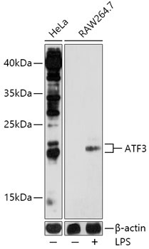
Western blot analysis of various lysates using ATF3 Rabbit pAb (CAB13469) at 1:1000 dilution. Raw264.7 cells were treated with LPS (1 μg/mL) at 37℃ for 8 hours. Secondary antibody: HRP-conjugated Goat anti-Rabbit IgG (H+L) (CABS014) at 1:10000 dilution. Lysates/proteins: 25μg per lane. Blocking buffer: 3% nonfat dry milk in TBST. Detection: ECL Basic Kit (AbGn00020). Exposure time: 120s.
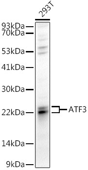
Western blot analysis of lysates from 293T cells, using ATF3 Rabbit pAb (CAB13469) at 1:1000 dilution. Secondary antibody: HRP-conjugated Goat anti-Rabbit IgG (H+L) (CABS014) at 1:10000 dilution. Lysates/proteins: 25μg per lane. Blocking buffer: 3% nonfat dry milk in TBST. Detection: ECL Basic Kit (AbGn00020). Exposure time: 30s.
Immunohistochemistry analysis of paraffin-embedded Human lung cancer using ATF3 Rabbit pAb (CAB13469) at dilution of 1:300 (40x lens). High pressure antigen retrieval performed with 0.01M Citrate buffer (pH 6.0) prior to IHC staining.