The ATF6 Antibody (CAB0202) is a high-quality antibody developed for reliable detection and analysis of target proteins. This antibody, produced in rabbits, exhibits high reactivity with human samples and is validated for use in Western blot applications. By specifically binding to ATF6, researchers can detect and analyze this important protein in a variety of cell types, making it ideal for studies in molecular biology and disease research.ATF6, also known as activating transcription factor 6, plays a crucial role in the cellular stress response by regulating the expression of genes involved in protein folding and quality control.
This antibody is validated for use in WB, IHC-P, IF/ICC, ELISA applications and has demonstrated reactivity against Human, Mouse, Rat samples.
Product Name:
ATF6 Antibody
SKU:
CAB0202
Size:
20μL, 100μL
Reactivity:
Human, Mouse, Rat
Conjugate:
Unconjugated
Immunogen:
Recombinant protein (or fragment).This information is considered to be commercially sensitive.
Recommended starting concentration is 1 μg/mL. Please optimize the concentration based on your specific assay requirements.
Synonyms:
ACHM7, ATF6A, ATF6
Positive Sample:
Rat lung
Cellular Localization:
Endoplasmic Reticulum Membrane, Nucleus, Single-Pass Type Ii Membrane Protein.
Calculated MW:
75kDa
Observed MW:
90-100kDa
This gene encodes a transcription factor that activates target genes for the unfolded protein response (UPR) during endoplasmic reticulum (ER) stress. Although it is a transcription factor, this protein is unusual in that it is synthesized as a transmembrane protein that is embedded in the ER. It functions as an ER stress sensor/transducer, and following ER stress-induced proteolysis, it functions as a nuclear transcription factor via a cis-acting ER stress response element (ERSE) that is present in the promoters of genes encoding ER chaperones. This protein has been identified as a survival factor for quiescent but not proliferative squamous carcinoma cells. There have been conflicting reports about the association of polymorphisms in this gene with diabetes in different populations, but another polymorphism has been associated with increased plasma cholesterol levels. This gene is also thought to be a potential therapeutic target for cystic fibrosis.
Purification Method
Affinity purification
Gene ID
22926
RRID
AB_2757016
Buffer Information
Store at -20℃. Avoid freeze / thaw cycles. Buffer: PBS containing 50% glycerol, preserved with proclin300 or sodium azide, pH 7.3.
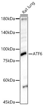
Western blot analysis of lysates from Rat lung using ATF6 Rabbit pAb (CAB0202) at 1:500 dilution. Secondary antibody: HRP-conjugated Goat anti-Rabbit IgG (H+L) (CABS014) at 1:10000 dilution. Lysates/proteins: 25 μg per lane. Blocking buffer: 3% nonfat dry milk in TBST. Detection: ECL Basic Kit (AbGn00020). Exposure time: 90s.
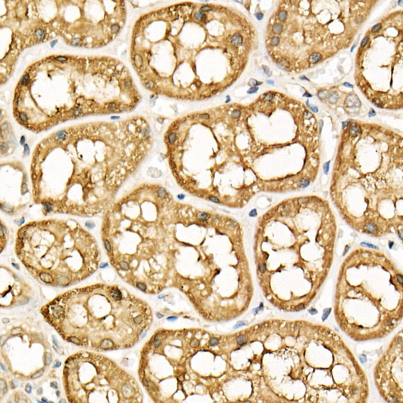
Immunohistochemistry analysis of paraffin-embedded Human kidney using ATF6 Rabbit pAb (CAB0202) at dilution of 1:20 (40x lens). High pressure antigen retrieval performed with 0.01M Citrate buffer (pH 6.0) prior to IHC staining.
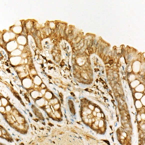
Immunohistochemistry analysis of paraffin-embedded Rat colon using ATF6 Rabbit pAb (CAB0202) at dilution of 1:20 (40x lens). High pressure antigen retrieval performed with 0.01M Citrate buffer (pH 6.0) prior to IHC staining.
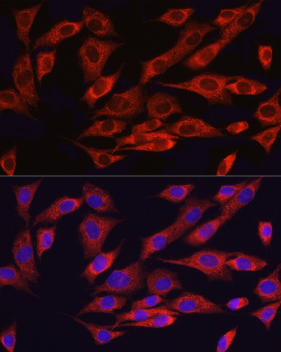
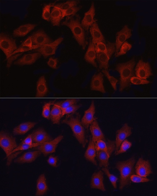
Immunofluorescence analysis of NIH/3T3 cells using ATF6 Rabbit pAb (CAB0202) at dilution of 1:50 (40x lens). Secondary antibody: Cy3-conjugated Goat anti-Rabbit IgG (H+L) (CABS007) at 1:500 dilution. Blue: DAPI for nuclear staining.
Immunofluorescence analysis of PC-12 cells using ATF6 Rabbit pAb (CAB0202) at dilution of 1:50 (40x lens). Secondary antibody: Cy3-conjugated Goat anti-Rabbit IgG (H+L) (CABS007) at 1:500 dilution. Blue: DAPI for nuclear staining.