The ATG4C Antibody (CAB7396) is a high-quality antibody developed for reliable detection and analysis of target proteins. This antibody, generated in rabbits, exhibits high specificity and sensitivity towards human samples, making it an ideal choice for Western blot applications. By binding to the ATG4C protein, this antibody allows for the detection and analysis of ATG4C in various cell types, offering insights into its role in autophagy regulation.ATG4C is a critical factor in the autophagy pathway, involved in the cleavage of the LC3 protein and the maturation of autophagosomes.
This antibody is validated for use in WB, ELISA applications and has demonstrated reactivity against Human samples.
Product Name:
ATG4C Antibody
SKU:
CAB7396
Size:
20μL, 100μL
Reactivity:
Human
Conjugate:
Unconjugated
Immunogen:
Recombinant protein (or fragment).This information is considered to be commercially sensitive.
Recommended starting concentration is 1 μg/mL. Please optimize the concentration based on your specific assay requirements.
Synonyms:
APG4C, AUTL1, AUTL3, APG4-C, HsAPG4C, ATG4C
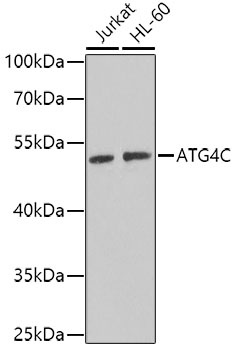
Positive Sample:
Jurkat, HL-60
Cellular Localization:
Cytoplasm.
Calculated MW:
52kDa
Observed MW:
52kDa
Autophagy is the process by which endogenous proteins and damaged organelles are destroyed intracellularly. Autophagy is postulated to be essential for cell homeostasis and cell remodeling during differentiation, metamorphosis, non-apoptotic cell death, and aging. Reduced levels of autophagy have been described in some malignant tumors, and a role for autophagy in controlling the unregulated cell growth linked to cancer has been proposed. This gene encodes a member of the autophagin protein family. The encoded protein is also designated as a member of the C-54 family of cysteine proteases. Alternate transcriptional splice variants, encoding the same protein, have been characterized.
Purification Method
Affinity purification
Gene ID
84938
RRID
AB_2767928
Buffer Information
Store at -20℃. Avoid freeze / thaw cycles. Buffer: PBS containing 50% glycerol, preserved with proclin300 or sodium azide, pH 7.3.
Western blot analysis of various lysates using ATG4C Rabbit pAb (CAB7396) at 1:1000 dilution. Secondary antibody: HRP-conjugated Goat anti-Rabbit IgG (H+L) (CABS014) at 1:10000 dilution. Lysates/proteins: 25μg per lane. Blocking buffer: 3% nonfat dry milk in TBST. Detection: ECL Basic Kit (AbGn00020). Exposure time: 90s.