The ATOH8 Antibody (CAB17232) is a high-quality antibody developed for reliable detection and analysis of target proteins. This antibody, produced in rabbits, has high specificity for human samples and is validated for use in various applications, including Western blotting.ATOH8, a transcription factor belonging to the basic helix-loop-helix (bHLH) family, is known to be involved in processes such as neurogenesis and cell differentiation. By specifically binding to ATOH8, this polyclonal antibody enables scientists to detect and study the function of this protein in different cell types, making it a valuable tool for research in developmental biology and related fields.
This antibody is validated for use in WB, ELISA applications and has demonstrated reactivity against Mouse, Rat samples.
Product Name:
ATOH8 Antibody
SKU:
CAB17232
Size:
20μL, 100μL
Reactivity:
Mouse, Rat
Immunogen:
Synthetic peptide. This information is considered to be commercially sensitive.
Sequence:
LRPR ILLC APPA RPAP SAPP APPA PPES TVRP APPT RPGE SSYS SISH VIYN NHQD SSAS PRKR PGEA TAAS SEIK ALQQ TRRL LANA RERT RVHT ISAA F
Tested Applications:
WBELISA
Recommended Dilution:
WB
1:500 - 1:2000
ELISA
Recommended starting concentration is 1 μg/mL. Please optimize the concentration based on your specific assay requirements.
Synonyms:
HATH6, bHLHa21, ATOH8
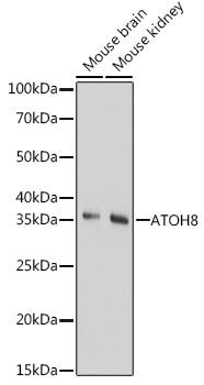
Positive Sample:
Mouse brain, Mouse kidney
Cellular Localization:
Cytoplasm, Nuclear Speck, Nucleoplasm, Nucleus.
Calculated MW:
35kDa
Observed MW:
35kDa
Enables DNA-binding transcription factor activity and E-box binding activity. Involved in several processes, including SMAD protein signal transduction; positive regulation of endothelial cell differentiation; and regulation of gene expression. Located in nucleoplasm.
Purification Method
Affinity purification
Gene ID
84913
RRID
AB_2768496
Buffer Information
Store at -20℃. Avoid freeze / thaw cycles. Buffer: PBS with 0.01% thimerosal,50% glycerol,pH7.3.
Western blot analysis of various lysates using ATOH8 Rabbit pAb (CAB17232) at 1:1000 dilution. Secondary antibody: HRP-conjugated Goat anti-Rabbit IgG (H+L) (CABS014) at 1:10000 dilution. Lysates/proteins: 25μg per lane. Blocking buffer: 3% nonfat dry milk in TBST. Detection: ECL Basic Kit (AbGn00020). Exposure time: 180s.