The ATP23 Antibody (CAB12885) is a high-quality antibody developed for reliable detection and analysis of target proteins. This antibody, produced in rabbits, exhibits high reactivity with human samples and has been validated for use in various applications, including Western blotting. By binding specifically to the ATP23 protein, researchers can accurately detect and analyze its expression in different cell types, making it an essential component for studies in mitochondrial biology and related diseases. ATP23, a member of the ATPase family, plays a crucial role in the maintenance of mitochondrial membrane integrity and function.
This antibody is validated for use in WB, IHC-P, IF/ICC, ELISA applications and has demonstrated reactivity against Human, Mouse, Rat samples.
Product Name:
ATP23 Antibody
SKU:
CAB12885
Size:
20μL, 100μL
Reactivity:
Human, Mouse, Rat
Conjugate:
Unconjugated
Immunogen:
Recombinant protein (or fragment).This information is considered to be commercially sensitive.
Recommended starting concentration is 1 μg/mL. Please optimize the concentration based on your specific assay requirements.
Synonyms:
KUB3, XRCC6BP1, ATP23
Positive Sample:
Mouse brain, Rat brain
Cellular Localization:
Cytosol, Extrinsic Component Of Mitochondrial Inner Membrane, Plasma Membrane.
Calculated MW:
28kDa
Observed MW:
28kDa
The protein encoded by this gene is amplified in glioblastomas and interacts with the DNA binding subunit of DNA-dependent protein kinase. This kinase is involved in double-strand break repair (DSB), and higher expression of the encoded protein increases the efficiency of DSB. In addition, comparison to orthologous proteins strongly suggests that this protein is a metalloprotease important in the biosynthesis of mitochondrial ATPase. Several transcript variants encoding different isoforms have been found for this gene.
Purification Method
Affinity purification
Gene ID
91419
RRID
AB_2759728
Buffer Information
Store at -20℃. Avoid freeze / thaw cycles. Buffer: Buffer: PBS containing 50% glycerol, preserved with proclin300 or sodium azide, pH 7.3.
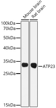
Western blot analysis of various lysates using ATP23 Rabbit pAb (CAB12885) at 1:3000 dilution. Secondary antibody: HRP-conjugated Goat anti-Rabbit IgG (H+L) (CABS014) at 1:10000 dilution. Lysates / proteins: 25 μg per lane. Blocking buffer: 3 % nonfat dry milk in TBST. Detection: ECL Basic Kit (AbGn00020). Exposure time: 90s.
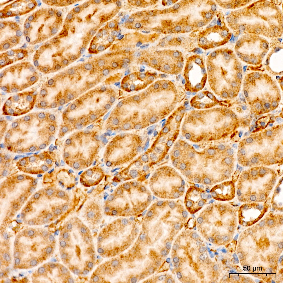
Immunohistochemistry analysis of paraffin-embedded Rat kidney tissue using ATP23 Rabbit pAb (CAB12885) at a dilution of 1:100 (40x lens). High pressure antigen retrieval was performed with 0.01 M citrate buffer (pH 6.0) prior to IHC staining.
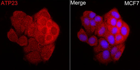
Immunofluorescence analysis of MCF7 cells using ATP23 Rabbit pAb (CAB12885) at dilution of 1:100 (40x lens). Secondary antibody: Cy3-conjugated Goat anti-Rabbit IgG (H+L) (CABS007) at 1:500 dilution. Blue: DAPI for nuclear staining.