The ATP5L Antibody (CAB9178) is a high-quality antibody developed for reliable detection and analysis of target proteins. Mitochondrial ATP synthase catalyzes ATP synthesis, utilizing an electrochemical gradient of protons across the inner membrane during oxidative phosphorylation. It is composed of two linked multi-subunit complexes: the soluble catalytic core, F1, and the membrane-spanning component, Fo, which comprises the proton channel. The F1 complex consists of 5 different subunits (alpha, beta, gamma, delta, and epsilon) assembled in a ratio of 3 alpha, 3 beta, and a single representative of the other 3. The Fo seems to have nine subunits (a, b, c, d, e, f, g, F6 and 8). This gene encodes the g subunit of the Fo complex. Alternative splicing results in multiple transcript variants.
This antibody is validated for use in WB, IF/ICC, ELISA applications and has demonstrated reactivity against Human, Mouse, Rat samples.
Product Name:
ATP5L Antibody
SKU:
CAB9178
Size:
100μL, 20μL
Reactivity:
Human, Mouse, Rat
Conjugate:
Unconjugated
Immunogen:
Recombinant protein (or fragment).This information is considered to be commercially sensitive.
Tested Applications:
WBIF/ICCELISA
Recommended Dilution:
WB
1:1000 - 1:5000
IF/ICC
1:50 - 1:200
ELISA
Recommended starting concentration is 1 μg/mL. Please optimize the concentration based on your specific assay requirements.
Synonyms:
ATP5L, ATP5JG
Positive Sample:
293T, A-549, HepG2, HeLa, Mouse brain, Mouse liver, Rat brain, Rat kidney
Cellular Localization:
Mitochondrion, Mitochondrion Inner Membrane.
Calculated MW:
11kDa
Observed MW:
11kDa
Mitochondrial ATP synthase catalyzes ATP synthesis, utilizing an electrochemical gradient of protons across the inner membrane during oxidative phosphorylation. It is composed of two linked multi-subunit complexes: the soluble catalytic core, F1, and the membrane-spanning component, Fo, which comprises the proton channel. The F1 complex consists of 5 different subunits (alpha, beta, gamma, delta, and epsilon) assembled in a ratio of 3 alpha, 3 beta, and a single representative of the other 3. The Fo seems to have nine subunits (a, b, c, d, e, f, g, F6 and 8). This gene encodes the g subunit of the Fo complex. Alternative splicing results in multiple transcript variants.
Purification Method
Affinity purification
Gene ID
10632
RRID
AB_2768508
Buffer Information
Store at -20℃. Avoid freeze / thaw cycles. Buffer: PBS with 0.01% thimerosal,50% glycerol,pH7.3.
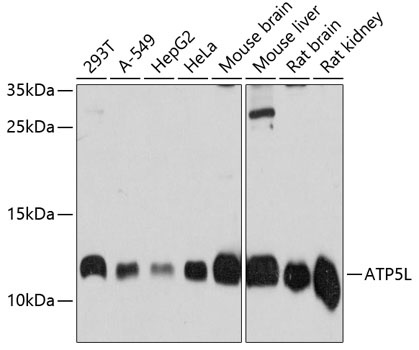
Western blot analysis of various lysates using ATP5L Rabbit pAb (CAB9178) at 1:3000 dilution. Secondary antibody: HRP-conjugated Goat anti-Rabbit IgG (H+L) (AS014) at 1:10000 dilution. Lysates/proteins: 25μg per lane. Blocking buffer: 3% nonfat dry milk in TBST. Detection: ECL Basic Kit (AbGn00020). Exposure time: 3s.
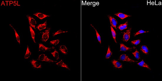
Immunofluorescence analysis of HeLa cells using ATP5L Rabbit pAb(CAB9178) at a dilution of 1:100 (40x lens). Secondary antibody:Cy3 Goat Anti-Rabbit IgG (H+L)(AS007) at 1:500 dilution. Blue: DAPI for nuclear staining.
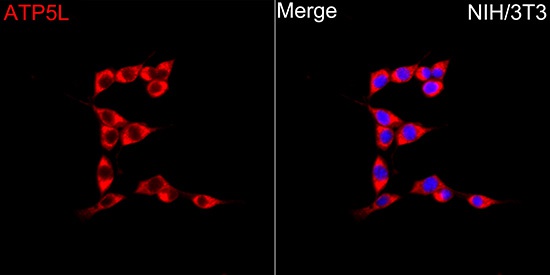
Immunofluorescence analysis of NIH/3T3 cells using ATP5L Rabbit pAb(CAB9178) at a dilution of 1:100 (40x lens). Secondary antibody:Cy3 Goat Anti-Rabbit IgG (H+L)(AS007) at 1:500 dilution. Blue: DAPI for nuclear staining.
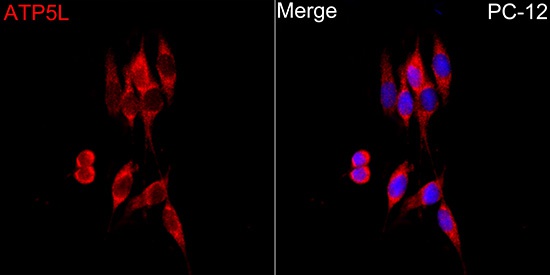
Immunofluorescence analysis of PC-12 cells using ATP5L Rabbit pAb(CAB9178) at a dilution of 1:100 (40x lens). Secondary antibody:Cy3 Goat Anti-Rabbit IgG (H+L)(AS007) at 1:500 dilution. Blue: DAPI for nuclear staining.


 ELISA Kit (HUFI03337)")

