The ATP6V1B2 Antibody (CAB3754) is a high-quality antibody developed for reliable detection and analysis of target proteins. The antibody, raised in rabbits, is validated for use in applications such as Western blot and immunohistochemistry, enabling precise detection and analysis of ATP6V1B2 expression in human samples.ATP6V1B2, a key component of the V-ATPase complex, plays a crucial role in regulating intracellular pH, membrane trafficking, and organelle acidification. Dysregulation of ATP6V1B2 expression has been implicated in various diseases, including cancer, osteopetrosis, and renal tubular acidosis.
This antibody is validated for use in WB, ELISA applications and has demonstrated reactivity against Human, Mouse, Rat samples.
Product Name:
ATP6V1B2 Antibody
SKU:
CAB3754
Size:
20μL, 100μL
Reactivity:
Human, Mouse, Rat
Conjugate:
Unconjugated
Immunogen:
Recombinant protein (or fragment).This information is considered to be commercially sensitive.
This gene encodes a component of vacuolar ATPase (V-ATPase), a multisubunit enzyme that mediates acidification of eukaryotic intracellular organelles. V-ATPase dependent organelle acidification is necessary for such intracellular processes as protein sorting, zymogen activation, receptor-mediated endocytosis, and synaptic vesicle proton gradient generation. V-ATPase is composed of a cytosolic V1 domain and a transmembrane V0 domain. The V1 domain consists of three A, three B, and two G subunits, as well as a C, D, E, F, and H subunit. The V1 domain contains the ATP catalytic site. The protein encoded by this gene is one of two V1 domain B subunit isoforms and is the only B isoform highly expressed in osteoclasts.
Purification Method
Affinity purification
Gene ID
526
RRID
AB_2765252
Buffer Information
Store at -20℃. Avoid freeze / thaw cycles. Buffer: PBS containing 50% glycerol, preserved with proclin300 or sodium azide, pH 7.3.
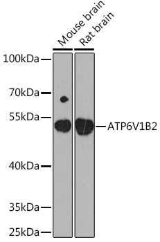
Western blot analysis of various lysates using ATP6V1B2 Rabbit pAb (CAB3754) at 1:1000 dilution. Secondary antibody: HRP-conjugated Goat anti-Rabbit IgG (H+L) (CABS014) at 1:10000 dilution. Lysates/proteins: 25μg per lane. Blocking buffer: 3% nonfat dry milk in TBST. Detection: ECL Basic Kit (AbGn00020). Exposure time: 30s.
Western blot analysis of various lysates using ATP6V1B2 Rabbit pAb (CAB3754) at 1:1000 dilution. Secondary antibody: HRP-conjugated Goat anti-Rabbit IgG (H+L) (CABS014) at 1:10000 dilution. Lysates/proteins: 25μg per lane. Blocking buffer: 3% nonfat dry milk in TBST. Detection: ECL Basic Kit (AbGn00020). Exposure time: 90s.