The ATP7B Antibody (CAB5676) is a high-quality antibody developed for reliable detection and analysis of target proteins. This antibody, raised in rabbits, is highly specific for human samples and is validated for use in various applications, including Western blotting. By binding to the ATP7B protein, this antibody enables the detection and analysis of ATP7B expression in different cell types, making it ideal for studies in biochemistry and neuroscience.ATP7B plays a crucial role in copper transport and metabolism, with mutations in the ATP7B gene leading to disorders such as Wilson's disease, a hereditary disorder resulting in copper accumulation and toxicity.
This antibody is validated for use in WB, IHC-P, ELISA applications and has demonstrated reactivity against Human, Mouse, Rat samples.
Product Name:
ATP7B Antibody
SKU:
CAB5676
Size:
20μL, 100μL
Reactivity:
Human, Mouse, Rat
Conjugate:
Unconjugated
Immunogen:
Recombinant protein (or fragment).This information is considered to be commercially sensitive.
This gene is a member of the P-type cation transport ATPase family and encodes a protein with several membrane-spanning domains, an ATPase consensus sequence, a hinge domain, a phosphorylation site, and at least 2 putative copper-binding sites. This protein is a monomer, and functions as a copper-transporting ATPase which exports copper out of the cells, such as the efflux of hepatic copper into the bile. Alternate transcriptional splice variants, encoding different isoforms with distinct cellular localizations, have been characterized. Mutations in this gene have been associated with Wilson disease which is characterized by copper accumulation.
Purification Method
Affinity purification
Gene ID
540
RRID
AB_2766436
Buffer Information
Store at -20℃. Avoid freeze / thaw cycles. Buffer: PBS with 0.09% Sodium azide,50% glycerol,pH7.3.
Western blot analysis of lysates from Rat liver using ATP7B Rabbit pAb (CAB5676) at 1:1000 dilution. Secondary antibody: HRP-conjugated Goat anti-Rabbit IgG (H+L) (CABS014) at 1:10000 dilution. Lysates/proteins: 25 μg per lane. Blocking buffer: 3% nonfat dry milk in TBST. Detection: ECL Basic Kit (AbGn00020). Exposure time: 180s.
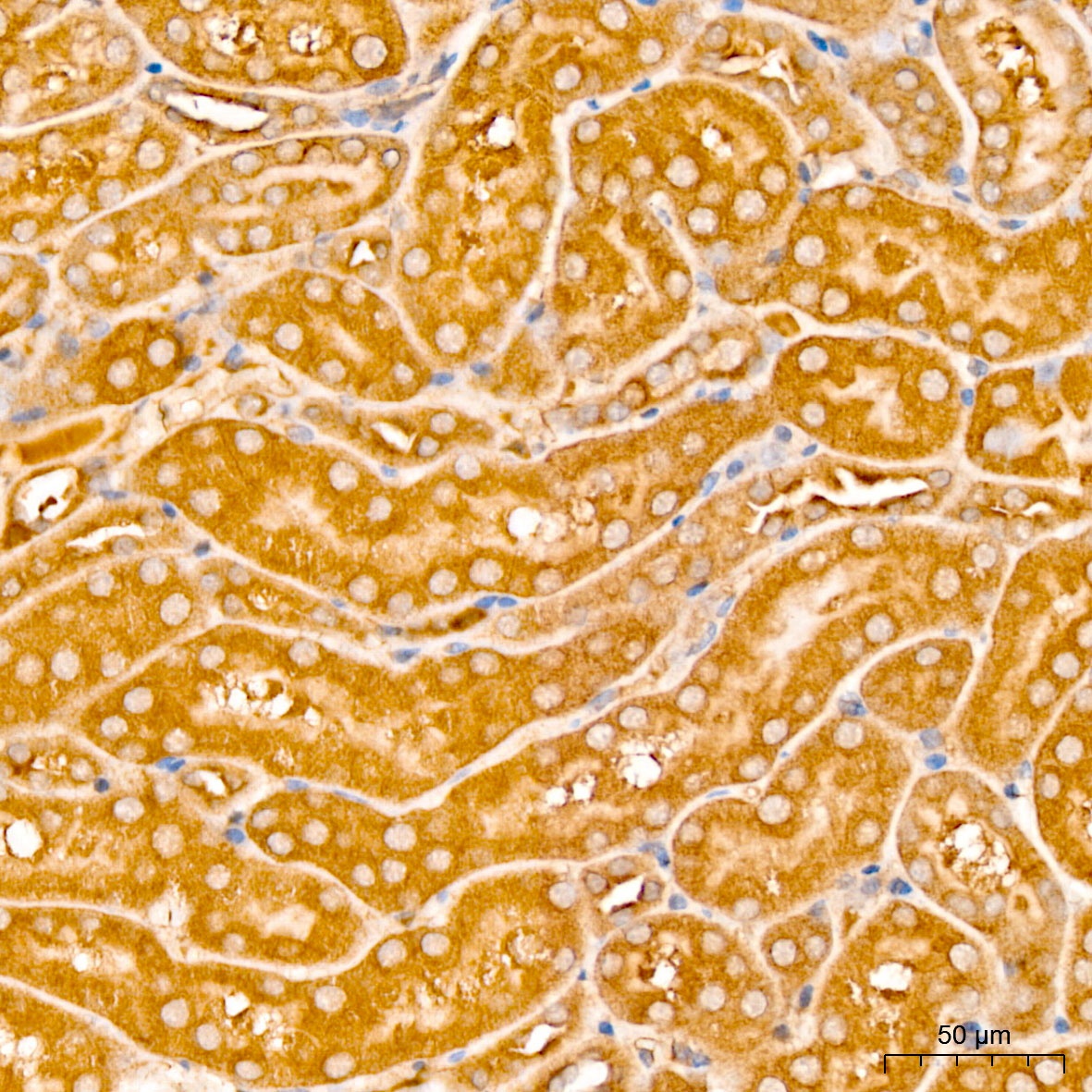
Immunohistochemistry analysis of paraffin-embedded Mouse kidney tissue using ATP7B Rabbit pAb (CAB5676) at a dilution of 1:100 (40x lens). High pressure antigen retrieval was performed with 0.01 M citrate buffer (pH 6.0) prior to IHC staining.
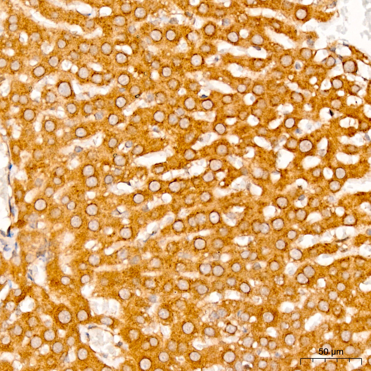
Immunohistochemistry analysis of paraffin-embedded Rat liver tissue using ATP7B Rabbit pAb (CAB5676) at a dilution of 1:100 (40x lens). High pressure antigen retrieval was performed with 0.01 M citrate buffer (pH 6.0) prior to IHC staining.
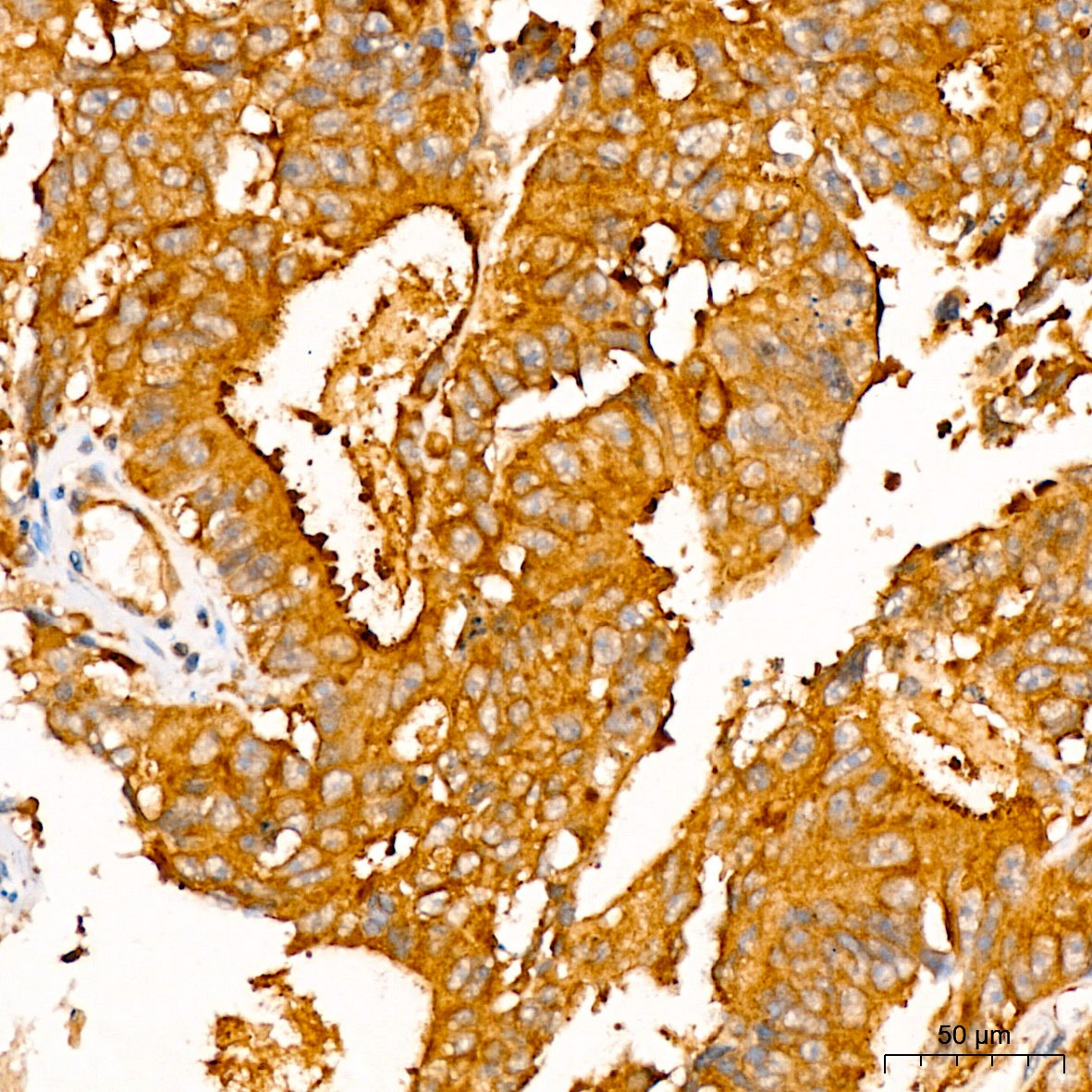
Immunohistochemistry analysis of paraffin-embedded Human colon carcinoma tissue using ATP7B Rabbit pAb (CAB5676) at a dilution of 1:100 (40x lens). High pressure antigen retrieval was performed with 0.01 M citrate buffer (pH 6.0) prior to IHC staining.