The Aurora B Monoclonal Antibody (CAB19539) is a high-quality antibody developed for reliable detection and analysis of target proteins. This gene encodes a member of the aurora kinase subfamily of serine/threonine kinases. The genes encoding the other two members of this subfamily are located on chromosomes 19 and 20. These kinases participate in the regulation of alignment and segregation of chromosomes during mitosis and meiosis through association with microtubules. A pseudogene of this gene is located on chromosome 8. Alternatively spliced transcript variants have been found for this gene.
This antibody is validated for use in WB, IF/ICC, ELISA applications and has demonstrated reactivity against Human, Mouse, Rat samples.
Product Name:
Aurora B Monoclonal Antibody
SKU:
CAB19539
Size:
100μL, 20μL
Reactivity:
Human, Mouse, Rat
Clone Number:
ARC50905
Conjugate:
Unconjugated
Immunogen:
Synthetic peptide. This information is considered to be commercially sensitive.
Tested Applications:
WBIF/ICCELISA
Recommended Dilution:
WB
1:1000 - 1:2000
IF/ICC
1:400 - 1:1600
ELISA
Recommended starting concentration is 1 μg/mL. Please optimize the concentration based on your specific assay requirements.
This gene encodes a member of the aurora kinase subfamily of serine/threonine kinases. The genes encoding the other two members of this subfamily are located on chromosomes 19 and 20. These kinases participate in the regulation of alignment and segregation of chromosomes during mitosis and meiosis through association with microtubules. A pseudogene of this gene is located on chromosome 8. Alternatively spliced transcript variants have been found for this gene.
Purification Method
Affinity purification
Gene ID
9212
RRID
AB_2862656
Buffer Information
Store at -20℃. Avoid freeze / thaw cycles. Buffer: PBS containing 50% glycerol and 0.05% BSA, preserved with proclin300 or sodium azide, pH 7.3.
Western blot analysis of various lysates using Aurora B Rabbit mAb (CAB19539) at 1:1000 dilution. Secondary antibody: HRP-conjugated Goat anti-Rabbit IgG (H+L) (AS014) at 1:10000 dilution. Lysates/proteins: 25μg per lane. Blocking buffer: 3% nonfat dry milk in TBST. Detection: ECL Basic Kit (AbGn00020). Exposure time: 3min.
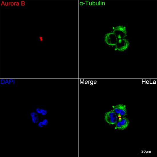
Confocal imaging of HeLa cells using Aurora B Rabbit mAb (CAB19539, dilution 1:800) followed by a further incubation with Cy3 Goat Anti-Rabbit IgG (H+L) (AS007, dilution 1:500) (Red). The cells were counterstained with α-Tubulin Mouse mAb (AC012, dilution 1:400) followed by incubation with ABflo® 488-conjugated Goat Anti-Mouse IgG (H+L) Ab (AS076, dilution 1:500) (Green). DAPI was used for nuclear staining (Blue). Objective: 100x.
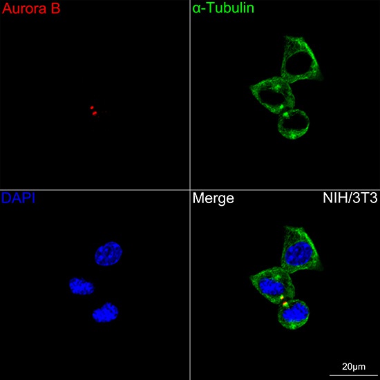
Confocal imaging of NIH/3T3 cells using Aurora B Rabbit mAb (CAB19539, dilution 1:800) followed by a further incubation with Cy3 Goat Anti-Rabbit IgG (H+L) (AS007, dilution 1:500) (Red). The cells were counterstained with α-Tubulin Mouse mAb (AC012, dilution 1:400) followed by incubation with ABflo® 488-conjugated Goat Anti-Mouse IgG (H+L) Ab (AS076, dilution 1:500) (Green). DAPI was used for nuclear staining (Blue). Objective: 100x.
Confocal imaging of C6 cells using Aurora B Rabbit mAb (CAB19539, dilution 1:800) followed by a further incubation with Cy3 Goat Anti-Rabbit IgG (H+L) (AS007, dilution 1:500) (Red). The cells were counterstained with α-Tubulin Mouse mAb (AC012, dilution 1:400) followed by incubation with ABflo® 488-conjugated Goat Anti-Mouse IgG (H+L) Ab (AS076, dilution 1:500) (Green). DAPI was used for nuclear staining (Blue). Objective: 100x.