The BAAT Antibody (CAB7646) is a high-quality antibody developed for reliable detection and analysis of target proteins. This antibody, generated in rabbits, offers high reactivity with human samples and has been validated for use in Western blot applications. By binding specifically to the BAAT protein, researchers can detect and analyze its expression in various cell types, making it an essential resource for studies in biochemistry and liver disease research.
This antibody is validated for use in WB, IHC-P, IF/ICC, ELISA applications and has demonstrated reactivity against Human, Mouse, Rat samples.
Product Name:
BAAT Antibody
SKU:
CAB7646
Size:
20μL, 100μL
Reactivity:
Human, Mouse, Rat
Conjugate:
Unconjugated
Immunogen:
Recombinant protein (or fragment).This information is considered to be commercially sensitive.
Recommended starting concentration is 1 μg/mL. Please optimize the concentration based on your specific assay requirements.
Synonyms:
BAT, HCHO, BACAT, BACD1, BAAT
Positive Sample:
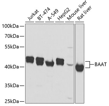
Jurkat, BT-474, A-549, HepG2, Mouse liver, Rat liver
Cellular Localization:
Cytoplasm.
Calculated MW:
46kDa
Observed MW:
40-46kDa
The protein encoded by this gene is a liver enzyme that catalyzes the transfer of C24 bile acids from the acyl-CoA thioester to either glycine or taurine, the second step in the formation of bile acid-amino acid conjugates. The bile acid conjugates then act as a detergent in the gastrointestinal tract, which enhances lipid and fat-soluble vitamin absorption. Defects in this gene are a cause of familial hypercholanemia (FHCA). Two transcript variants encoding the same protein have been found for this gene.
Purification Method
Affinity purification
Gene ID
570
RRID
AB_2768550
Buffer Information
Store at -20℃. Avoid freeze / thaw cycles. Buffer: PBS containing 50% glycerol, preserved with proclin300 or sodium azide, pH 7.3.
Western blot analysis of various lysates using BAAT Rabbit pAb (CAB7646) at 1:2000 dilution. Secondary antibody: HRP-conjugated Goat anti-Rabbit IgG (H+L) (CABS014) at 1:10000 dilution. Lysates/proteins: 25μg per lane. Blocking buffer: 3% nonfat dry milk in TBST. Detection: ECL Basic Kit (AbGn00020). Exposure time: 1s.
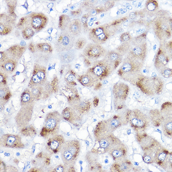
Immunohistochemistry analysis of paraffin-embedded Human liver using BAAT Rabbit pAb (CAB7646) at dilution of 1:500 (40x lens). High pressure antigen retrieval performed with 0.01M Citrate buffer (pH 6.0) prior to IHC staining.