The PUMA Antibody (CAB17138) is a high-quality antibody developed for reliable detection and analysis of target proteins. This antibody, generated in rabbits, exhibits high reactivity with human samples and has been validated for use in Western blotting applications.The BBC3 protein, also known as PUMA (p53 upregulated modulator of apoptosis), is involved in the regulation of cell death pathways and is a critical mediator of p53-induced apoptosis. Its role in promoting cell death in response to cellular stress makes it an important target for understanding the mechanisms of cancer development and therapy resistance.
This antibody is validated for use in WB, IHC-P, IF/ICC, ELISA applications and has demonstrated reactivity against Human, Mouse, Rat samples.
Product Name:
PUMA Antibody
SKU:
CAB17138
Size:
20μL, 100μL
Reactivity:
Human, Mouse, Rat
Immunogen:
Synthetic peptide. This information is considered to be commercially sensitive.
Recommended starting concentration is 1 μg/mL. Please optimize the concentration based on your specific assay requirements.
Synonyms:
JFY1, PUMA, JFY-1
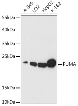
Positive Sample:
A-549, LO2, HepG2, K562
Cellular Localization:
Mitochondrion.
Calculated MW:
10kDa/14kDa/20kDa/26kDa
Observed MW:
20kDa
This gene encodes a member of the BCL-2 family of proteins. This family member belongs to the BH3-only pro-apoptotic subclass. The protein cooperates with direct activator proteins to induce mitochondrial outer membrane permeabilization and apoptosis. It can bind to anti-apoptotic Bcl-2 family members to induce mitochondrial dysfunction and caspase activation. Because of its pro-apoptotic role, this gene is a potential drug target for cancer therapy and for tissue injury. Alternative splicing results in multiple transcript variants.
Purification Method
Affinity purification
Gene ID
27113
RRID
AB_2768566
Buffer Information
Store at -20℃. Avoid freeze / thaw cycles. Buffer: PBS with 0.01% thimerosal,50% glycerol,pH7.3.
Western blot analysis of various lysates using PUMA Rabbit pAb (CAB17138) at 1:1000 dilution. Secondary antibody: HRP-conjugated Goat anti-Rabbit IgG (H+L) (CABS014) at 1:10000 dilution. Lysates/proteins: 25μg per lane. Blocking buffer: 3% nonfat dry milk in TBST. Detection: ECL Basic Kit (AbGn00020). Exposure time: 30s.
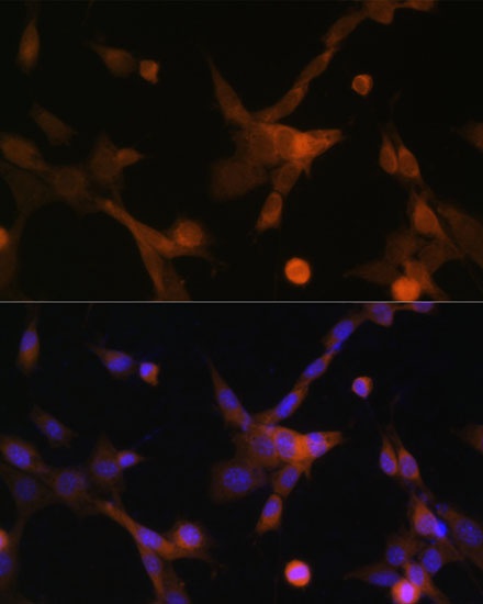
Immunofluorescence analysis of NIH-3T3 cells using PUMA Rabbit pAb (CAB17138) at dilution of 1:100 (40x lens). Secondary antibody: Cy3-conjugated Goat anti-Rabbit IgG (H+L) (CABS007) at 1:500 dilution. Blue: DAPI for nuclear staining.