The BCAT1 Antibody (CAB16351) is a high-quality antibody developed for reliable detection and analysis of target proteins. This antibody, generated in rabbits, exhibits strong reactivity with human samples and has been validated for use in Western blot applications. By specifically binding to the BCAT1 protein, this antibody allows for the detection and analysis of BCAT1 expression in a variety of cell types, making it an excellent tool for studies in metabolism and cancer research.BCAT1 plays a crucial role in regulating cellular metabolism by catalyzing the first step in the breakdown of branched-chain amino acids, which are essential for protein synthesis and energy production. Dysregulation of BCAT1 expression has been linked to various diseases, including cancer, metabolic disorders, and neurological conditions.
This antibody is validated for use in WB, IF/ICC, ELISA applications and has demonstrated reactivity against Human, Mouse, Rat samples.
Product Name:
BCAT1 Antibody
SKU:
CAB16351
Size:
20μL, 100μL
Reactivity:
Human, Mouse, Rat
Conjugate:
Unconjugated
Immunogen:
Recombinant protein (or fragment).This information is considered to be commercially sensitive.
Recommended starting concentration is 1 μg/mL. Please optimize the concentration based on your specific assay requirements.
Synonyms:
BCT1, PP18, BCATC, ECA39, MECA39, PNAS121, BCAT1
Positive Sample:
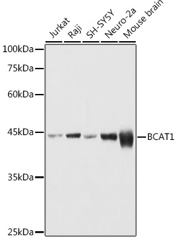
Jurkat, Raji, SH-SY5Y, Neuro-2a, Mouse brain
Cellular Localization:
Cytoplasm.
Calculated MW:
43kDa
Observed MW:
43kDa
This gene encodes the cytosolic form of the enzyme branched-chain amino acid transaminase. This enzyme catalyzes the reversible transamination of branched-chain alpha-keto acids to branched-chain L-amino acids essential for cell growth. Two different clinical disorders have been attributed to a defect of branched-chain amino acid transamination: hypervalinemia and hyperleucine-isoleucinemia. As there is also a gene encoding a mitochondrial form of this enzyme, mutations in either gene may contribute to these disorders. Alternatively spliced transcript variants have been described.
Purification Method
Affinity purification
Gene ID
586
RRID
AB_2768572
Buffer Information
Store at -20℃. Avoid freeze / thaw cycles. Buffer: PBS containing 50% glycerol, preserved with proclin300 or sodium azide, pH 7.3.
Western blot analysis of various lysates using BCAT1 Rabbit pAb (CAB16351) at 1:1000 dilution. Secondary antibody: HRP-conjugated Goat anti-Rabbit IgG (H+L) (CABS014) at 1:10000 dilution. Lysates/proteins: 25μg per lane. Blocking buffer: 3% nonfat dry milk in TBST. Detection: ECL Basic Kit (AbGn00020). Exposure time: 1s.
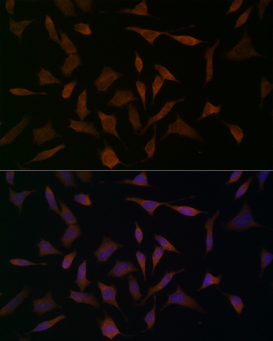
Immunofluorescence analysis of HeLa cells using BCAT1 Rabbit pAb (CAB16351) at dilution of 1:100 (40x lens). Secondary antibody: Cy3-conjugated Goat anti-Rabbit IgG (H+L) (CABS007) at 1:500 dilution. Blue: DAPI for nuclear staining.
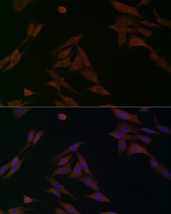
Immunofluorescence analysis of NIH/3T3 cells using BCAT1 Rabbit pAb (CAB16351) at dilution of 1:100 (40x lens). Secondary antibody: Cy3-conjugated Goat anti-Rabbit IgG (H+L) (CABS007) at 1:500 dilution. Blue: DAPI for nuclear staining.
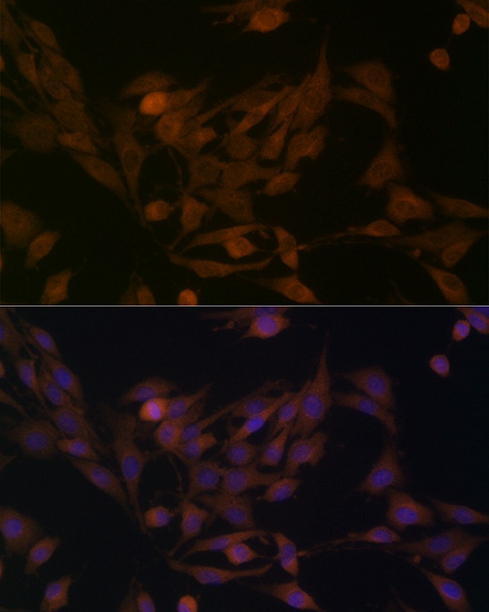
Immunofluorescence analysis of PC-12 cells using BCAT1 Rabbit pAb (CAB16351) at dilution of 1:100 (40x lens). Secondary antibody: Cy3-conjugated Goat anti-Rabbit IgG (H+L) (CABS007) at 1:500 dilution. Blue: DAPI for nuclear staining.