The BCL3 Antibody (CAB11582) is a high-quality antibody developed for reliable detection and analysis of target proteins. This antibody, generated in rabbits, exhibits high reactivity with human samples and is validated for use in Western blot applications. By binding specifically to the BCL3 protein, this antibody enables precise detection and analysis in various cell types, making it an essential component for studies in immunology and cancer research.
This antibody is validated for use in WB, IF/ICC, ELISA applications and has demonstrated reactivity against Human, Mouse, Rat samples.
Product Name:
BCL3 Antibody
SKU:
CAB11582
Size:
20μL, 100μL
Reactivity:
Human, Mouse, Rat
Conjugate:
Unconjugated
Immunogen:
Synthetic peptide. This information is considered to be commercially sensitive.
Sequence:
ANVN AQMY SGSS ALHS ASGR GLLP LVRT LVRS GADS SLKN CHND TPLM VARS RRVI DILR GKAT RPAS TSQP DPSP DRSA NTSP ESSS RLSS NGLL SASP S
Tested Applications:
WBIF/ICCELISA
Recommended Dilution:
WB
1:500 - 1:1000
IF/ICC
1:50 - 1:200
ELISA
Recommended starting concentration is 1 μg/mL. Please optimize the concentration based on your specific assay requirements.
Synonyms:
BCL4, D19S37, BCL3
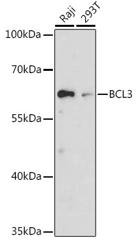
Positive Sample:
Raji, 293T
Cellular Localization:
Cytoplasm, Nucleus, Perinuclear Region.
Calculated MW:
48kDa
Observed MW:
60kDa
This gene is a proto-oncogene candidate. It is identified by its translocation into the immunoglobulin alpha-locus in some cases of B-cell leukemia. The protein encoded by this gene contains seven ankyrin repeats, which are most closely related to those found in I kappa B proteins. This protein functions as a transcriptional co-activator that activates through its association with NF-kappa B homodimers. The expression of this gene can be induced by NF-kappa B, which forms a part of the autoregulatory loop that controls the nuclear residence of p50 NF-kappa B.
Purification Method
Affinity purification
Gene ID
602
RRID
AB_2758612
Buffer Information
Store at -20℃. Avoid freeze / thaw cycles. Buffer: PBS with 0.01% thimerosal,50% glycerol,pH7.3.
Western blot analysis of various lysates using BCL3 Rabbit pAb (CAB11582) at 1:1000 dilution. Secondary antibody: HRP-conjugated Goat anti-Rabbit IgG (H+L) (CABS014) at 1:10000 dilution. Lysates/proteins: 25μg per lane. Blocking buffer: 3% nonfat dry milk in TBST. Detection: ECL Basic Kit (AbGn00020). Exposure time: 30s.
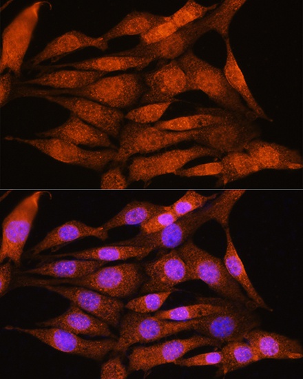
Immunofluorescence analysis of NIH/3T3 cells using BCL3 Rabbit pAb (CAB11582) at dilution of 1:100. Secondary antibody: Cy3-conjugated Goat anti-Rabbit IgG (H+L) (CABS007) at 1:500 dilution. Blue: DAPI for nuclear staining.
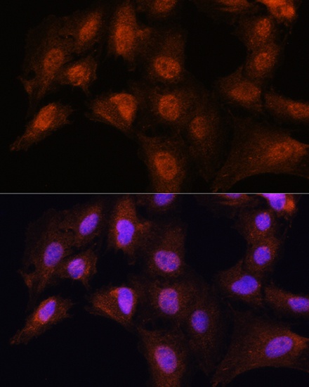
Immunofluorescence analysis of C6 cells using BCL3 Rabbit pAb (CAB11582) at dilution of 1:100. Secondary antibody: Cy3-conjugated Goat anti-Rabbit IgG (H+L) (CABS007) at 1:500 dilution. Blue: DAPI for nuclear staining.