The betaIII-Tubulin Monoclonal Antibody (CAB17913) is a high-quality antibody developed for reliable detection and analysis of target proteins. This gene encodes a class III member of the beta tubulin protein family. Beta tubulins are one of two core protein families (alpha and beta tubulins) that heterodimerize and assemble to form microtubules. This protein is primarily expressed in neurons and may be involved in neurogenesis and axon guidance and maintenance. Mutations in this gene are the cause of congenital fibrosis of the extraocular muscles type 3. Alternate splicing results in multiple transcript variants. A pseudogene of this gene is found on chromosome 6.
This antibody is validated for use in WB, IHC-P, IF/ICC, ELISA, IF-P applications and has demonstrated reactivity against Human, Mouse, Rat samples.
Product Name:
betaIII-Tubulin Monoclonal Antibody
SKU:
CAB17913
Size:
100μL, 20μL
Reactivity:
Human, Mouse, Rat
Clone Number:
ARC0456
Conjugate:
Unconjugated
Immunogen:
Synthetic peptide. This information is considered to be commercially sensitive.
Tested Applications:
WBIHC-PIF/ICCELISAIF-P
Recommended Dilution:
WB
1:10000 - 1:100000
IF/ICC
1:200 - 1:2000
IF-P
1:200 - 1:2000
IHC-P
1:200 - 1:4000
ELISA
Recommended starting concentration is 1 μg/mL. Please optimize the concentration based on your specific assay requirements.
SH-SY5Y, Mouse testis, Mouse brain, Rat testis, Rat brain
Cellular Localization:
Cytoplasm, Cytoskeleton.
Calculated MW:
50kDa
Observed MW:
55kDa
This gene encodes a class III member of the beta tubulin protein family. Beta tubulins are one of two core protein families (alpha and beta tubulins) that heterodimerize and assemble to form microtubules. This protein is primarily expressed in neurons and may be involved in neurogenesis and axon guidance and maintenance. Mutations in this gene are the cause of congenital fibrosis of the extraocular muscles type 3. Alternate splicing results in multiple transcript variants. A pseudogene of this gene is found on chromosome 6.
Purification Method
Affinity purification
Gene ID
10381
RRID
AB_2861758
Buffer Information
Store at -20℃. Avoid freeze / thaw cycles. Buffer: PBS containing 50% glycerol and 0.05% BSA, preserved with proclin300 or sodium azide, pH 7.3.
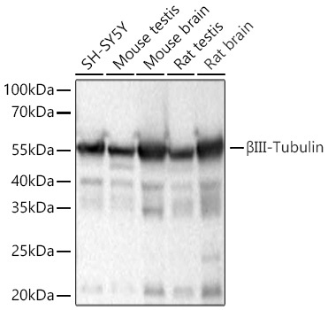
Western blot analysis of various lysates using βIII-Tubulin Rabbit mAb (CAB17913) at 1:50000 dilution incubated at room temperature for 1.5 hours. Secondary antibody: HRP-conjugated Goat anti-Rabbit IgG (H+L) (AS014) at 1:10000 dilution. Lysates/proteins: 25 μg per lane. Blocking buffer: 3% nonfat dry milk in TBST. Detection: ECL Basic Kit (AbGn00020). Exposure time: 45s.
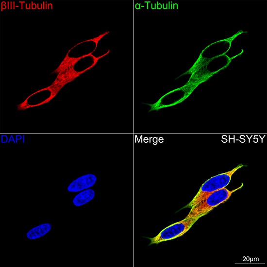
Confocal imaging of SH-SY5Y cells using βIII-Tubulin Rabbit mAb (CAB17913, dilution 1:200) followed by a further incubation with Cy3 Goat Anti-Rabbit IgG (H+L) (AS007, dilution 1:500) (Red). The cells were counterstained with α-Tubulin Mouse mAb (AC012, dilution 1:400) followed by incubation with ABflo® 488-conjugated Goat Anti-Mouse IgG (H+L) Ab (AS076, dilution 1:500) (Green). DAPI was used for nuclear staining (Blue). Objective: 100x.
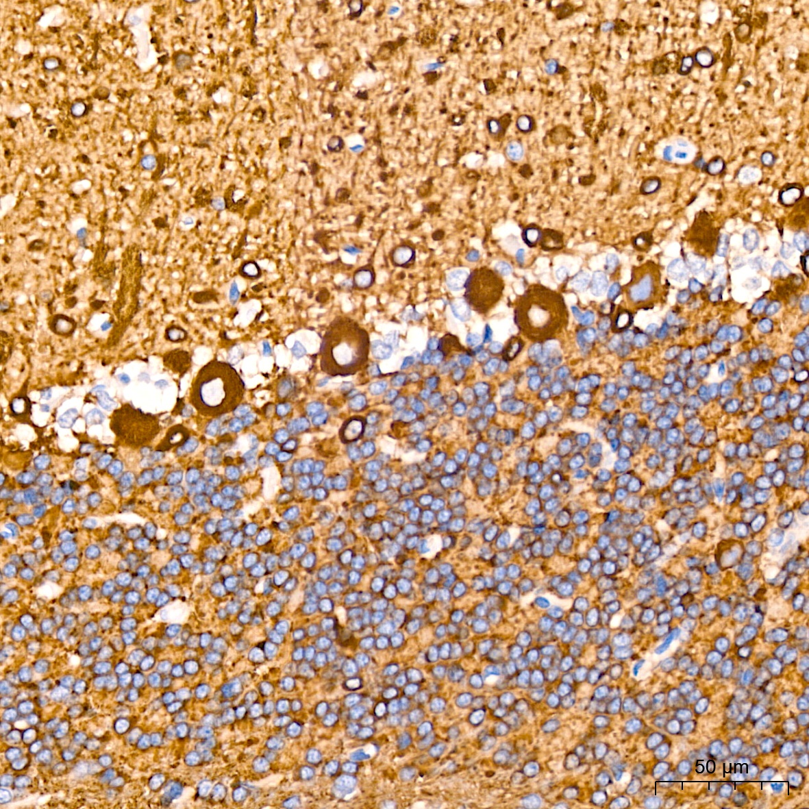
Immunohistochemistry analysis of paraffin-embedded Rat brain tissue using βIII-Tubulin Rabbit mAb (CAB17913) at a dilution of 1:200 (40x lens). High pressure antigen retrieval performed with 0.01M Citrate buffer (pH 6.0) prior to IHC staining.
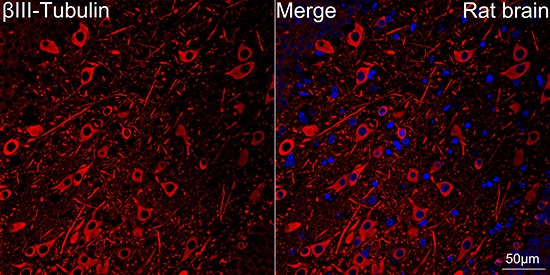
Confocal imaging of paraffin-embedded Rat brain tissue using βIII-Tubulin Rabbit mAb (CAB17913, dilution 1:200) followed by a further incubation with Cy3 Goat Anti-Rabbit IgG (H+L) (AS007, dilution 1:500) (Red). DAPI was used for nuclear staining (Blue). Objective: 40x. Perform microwave antigen retrieval with 0.01M citrate buffer (pH 6.0) prior to IF staining.