The cIAP2 Antibody (CAB0833) is a high-quality antibody developed for reliable detection and analysis of target proteins. This antibody, produced in rabbits, is highly specific for human samples and has been validated for use in Western blot applications. By binding to the BIRC3 protein, this antibody allows for precise detection and analysis of BIRC3 expression in a variety of cell types.BIRC3, also known as cIAP2, is a crucial player in cell signaling pathways that control apoptosis and inflammation. Dysregulation of BIRC3 has been implicated in various diseases, including cancer and autoimmune disorders.
This antibody is validated for use in WB, IHC-P, IF/ICC, ELISA applications and has demonstrated reactivity against Human, Mouse, Rat samples.
Product Name:
cIAP2 Antibody
SKU:
CAB0833
Size:
20μL, 100μL
Reactivity:
Human, Mouse, Rat
Conjugate:
Unconjugated
Immunogen:
Recombinant protein (or fragment).This information is considered to be commercially sensitive.
Sequence:
NSVN NLEA TSQP TFPS SVTN STHS LLPG TEN
Tested Applications:
WBIHC-PIF/ICCELISA
Recommended Dilution:
WB
1:100 - 1:500
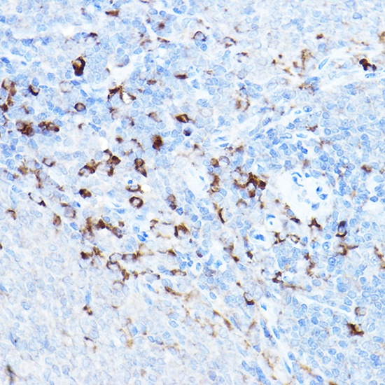
IHC-P
1:50 - 1:200
IF/ICC
1:50 - 1:200
ELISA
Recommended starting concentration is 1 μg/mL. Please optimize the concentration based on your specific assay requirements.
This gene encodes a member of the IAP family of proteins that inhibit apoptosis by binding to tumor necrosis factor receptor-associated factors TRAF1 and TRAF2, probably by interfering with activation of ICE-like proteases. The encoded protein inhibits apoptosis induced by serum deprivation but does not affect apoptosis resulting from exposure to menadione, a potent inducer of free radicals. It contains 3 baculovirus IAP repeats and a ring finger domain. Transcript variants encoding the same isoform have been identified.
Purification Method
Affinity purification
Gene ID
330
RRID
AB_2757419
Buffer Information
Store at -20℃. Avoid freeze / thaw cycles. Buffer: PBS containing 50% glycerol, preserved with proclin300 or sodium azide,pH7.3.
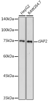
Western blot analysis of various lysates using cIAP2 Rabbit pAb (CAB0833) at 1:500 dilution. Secondary antibody: HRP-conjugated Goat anti-Rabbit IgG (H+L) (CABS014) at 1:10000 dilution. Lysates/proteins: 25μg per lane. Blocking buffer: 3% nonfat dry milk in TBST. Detection: ECL Basic Kit (AbGn00020). Exposure time: 180s.
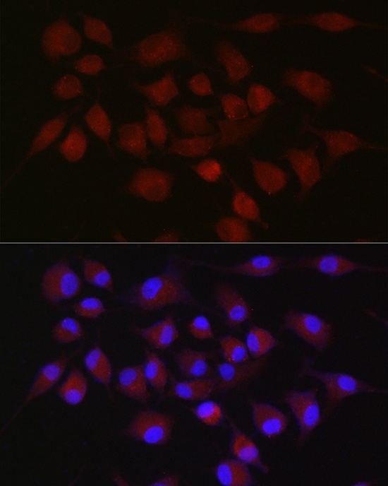
Immunofluorescence analysis of A-549 cells using cIAP2 Rabbit pAb (CAB0833) at dilution of 1:50 (40x lens). Secondary antibody: Cy3-conjugated Goat anti-Rabbit IgG (H+L) (CABS007) at 1:500 dilution. Blue: DAPI for nuclear staining.
Immunofluorescence analysis of A-549 cells using cIAP2 Rabbit pAb (CAB0833) at dilution of 1:50 (40x lens). Secondary antibody: Cy3-conjugated Goat anti-Rabbit IgG (H+L) (CABS007) at 1:500 dilution. Blue: DAPI for nuclear staining.
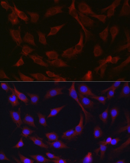
Immunofluorescence analysis of C6 cells using cIAP2 Rabbit pAb (CAB0833) at dilution of 1:50 (40x lens). Secondary antibody: Cy3-conjugated Goat anti-Rabbit IgG (H+L) (CABS007) at 1:500 dilution. Blue: DAPI for nuclear staining.