The BLID Antibody (CAB7275) is a high-quality antibody developed for reliable detection and analysis of target proteins. This gene encodes a BH3-like motif containing protein involved in cell death. The encoded protein may induce apoptosis in a caspase-dependent manner. The protein is localized in both the cytoplasm and the mitochondrion. RRID AB_2767816 Gene ID 414899 Swiss Prot Synonym BRCC2; BRCC2/BLID
This antibody is validated for use in WB, IF/ICC, ELISA applications and has demonstrated reactivity against Human, Mouse, Rat samples.
Product Name:
BLID Antibody
SKU:
CAB7275
Size:
100μL, 20μL
Reactivity:
Human, Mouse, Rat
Clone Number:
-
Conjugate:
Unconjugated
Immunogen:
Recombinant protein (or fragment).This information is considered to be commercially sensitive.
Tested Applications:
WBIF/ICCELISA
Recommended Dilution:
WB
1:500 - 1:1000
IF
/
ICC
1:50 - 1:100
ELISA
Recommended starting concentration is 1 μg/mL. Please optimize the concentration based on your specific assay requirements.
Synonyms:
BRCC2, BRCC2/BLID
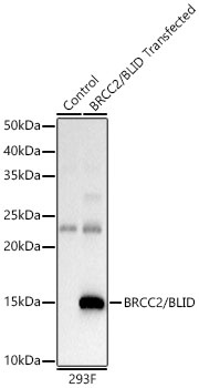
Positive Sample:
293T transfected with BRCC2/BLID
Cellular Localization:
Cytoplasm, Mitochondrion.
Calculated MW:
12kDa
Observed MW:
16kDa
This gene encodes a BH3-like motif containing protein involved in cell death. The encoded protein may induce apoptosis in a caspase-dependent manner. The protein is localized in both the cytoplasm and the mitochondrion. RRID AB_2767816 Gene ID 414899 Swiss Prot Synonym BRCC2; BRCC2/BLID
Purification Method:
Affinity purification
Gene ID:
414899
RRID:
AB_2767816
Buffer Information:
Store at -20℃. Avoid freeze / thaw cycles. Buffer: PBS containing 50% glycerol, preserved with proclin300 or sodium azide, pH 7.3.
Western blot analysis of lysates from wild type (WT) and 293T cells transfected with BRCC2/BLID, using BRCC2/BLID Rabbit pAb (CAB7275) at 1:400 dilution. Secondary antibody: HRP-conjugated Goat anti-Rabbit IgG (H+L) (AS014) at 1:10000 dilution. Lysates/proteins: 25μg per lane. Blocking buffer: 3% nonfat dry milk in TBST. Detection: ECL Basic Kit (AbGn00020). Exposure time: 1s.
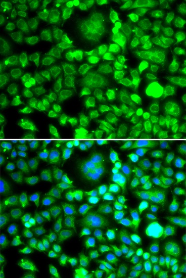
Immunofluorescence analysis of MCF-7 cells using BRCC2/BLID Rabbit pAb (CAB7275). Secondary antibody: Cy3-conjugated Goat anti-Rabbit IgG (H+L) (AS007) at 1:500 dilution. Blue: DAPI for nuclear staining.